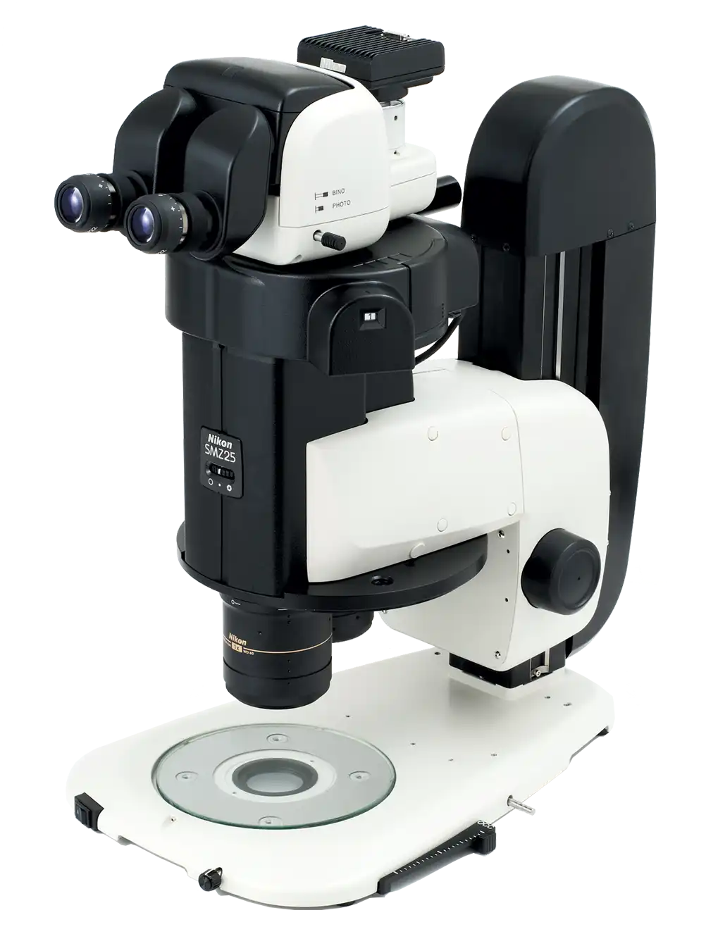
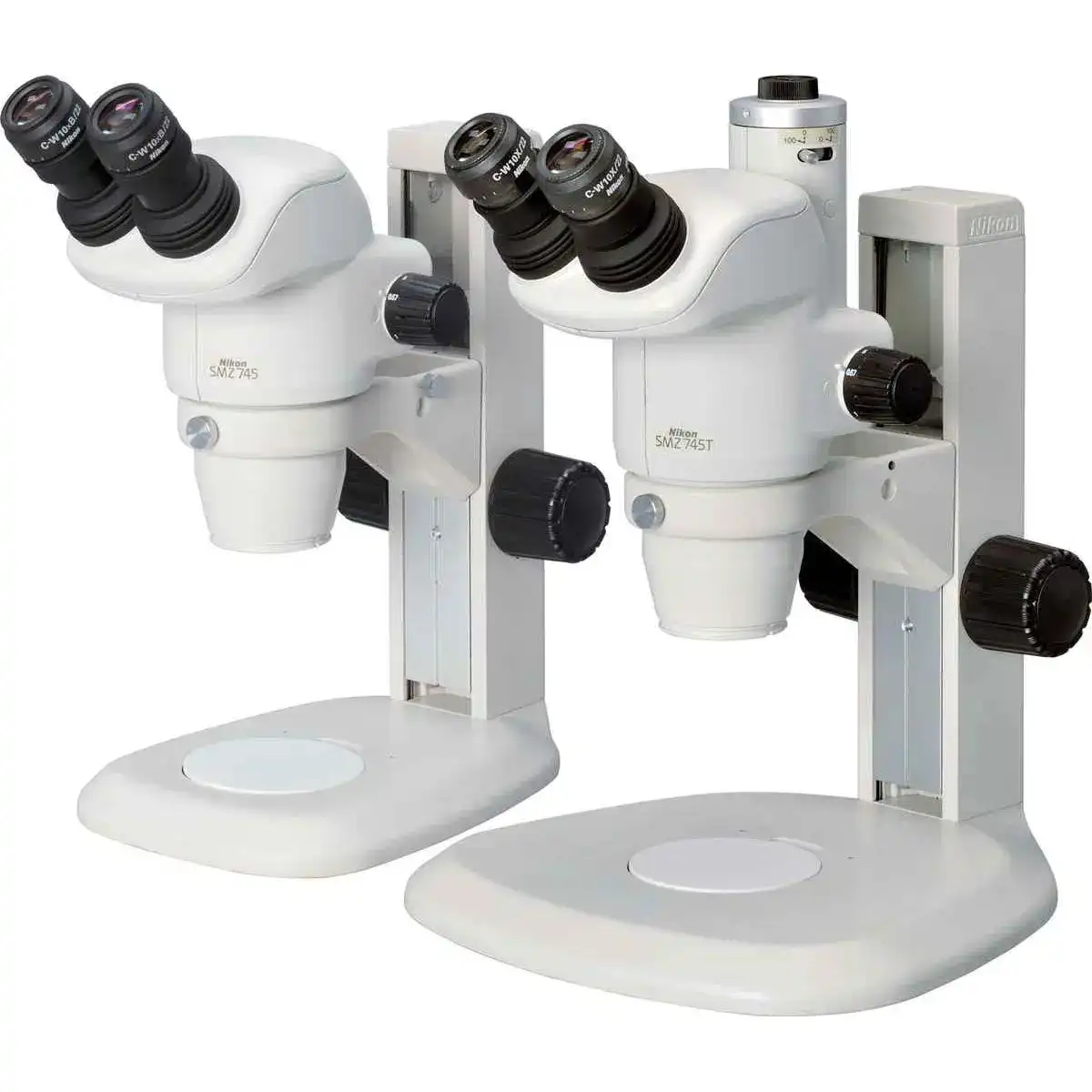
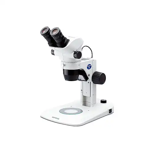
NIKON SMZ25,SMZ18
Research Stereomicroscopes
Breakthrough Stereo Microscope Optics
Traditional boundaries between scientific fields such as molecular biology and developmental biology are rapidly disappearing as researchers seek to connect findings at the molecular level to those derived from cellular, tissue, and organismal studies. Fields including molecular biology, cell biology, neurobiology, embryology, developmental biology and systems biology have increasing needs for imaging systems that span spatial scales from single cells to whole organisms.
With these demands in mind, Nikon has developed a stereo microscope that features a large zoom ratio of 25:1, high resolution and exceptional fluorescence transmission capability.
Key Features
Large zoom range enables high resolution macro to micro imaging
An innovative optical system known as Perfect Zoom System makes it possible for the first time to achieve a zoom ratio of 25:1 (zoom range: 0.63X - 15.75X). Even with a 1X objective lens, the SMZ25 captures the entire 35mm dish and simultaneously delivers microscopic details.
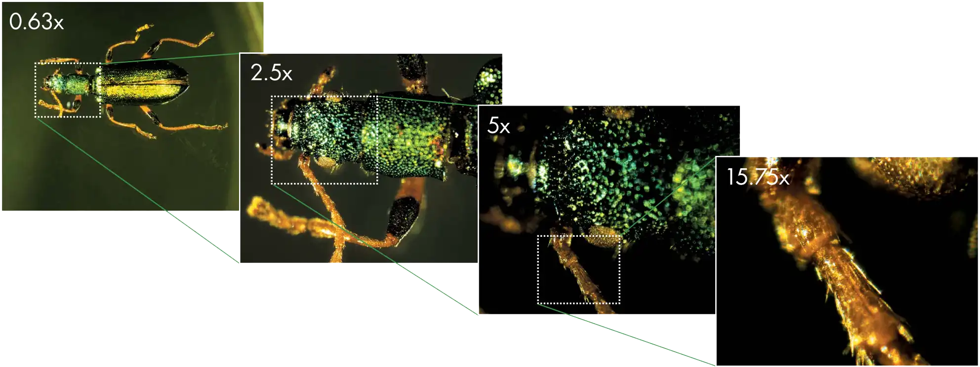
Arthromacra sp.
(Using SHR Plan Apo 1X with SMZ25)
Image courtesy of Japan Insect Association
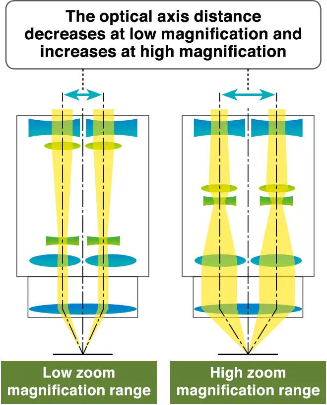
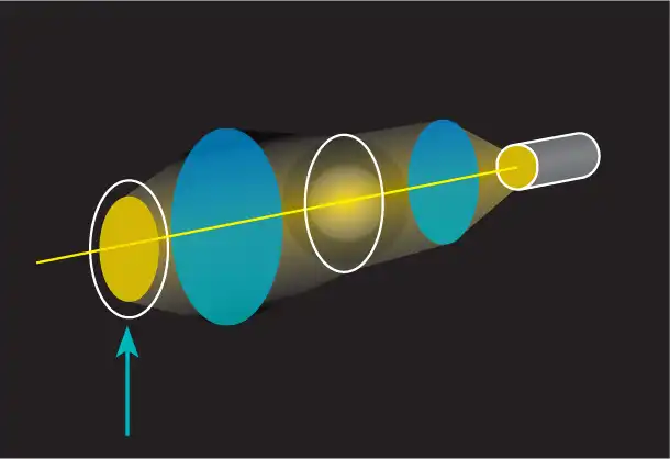
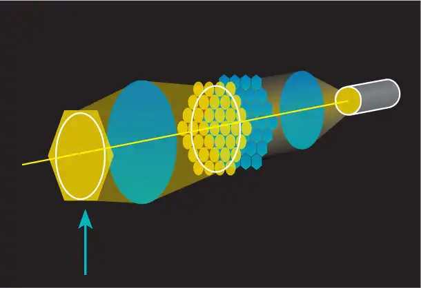
Nikon Perfect Zoom System offers new levels of imaging power and versatility
A breakthrough in stereo microscope design, Perfect Zoom System dynamically changes the distance between the two optical axes as the zoom factor is changed. This change in optical axis distance enables maximization of light entry into the optical system at every magnification. The result is an uncompromised, large zoom range, high resolution in both eye paths, and minimal aberrations over the entire zoom-range. Furthermore, this breakthrough in optical design enables all of these desirable features be housed in a compact zoom body, resulting in an ergonomic instrument design.
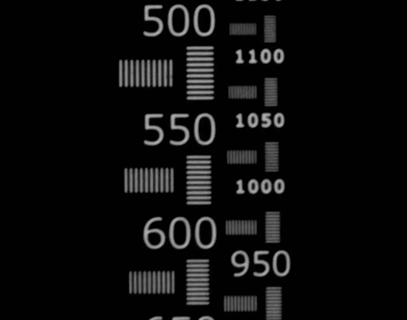
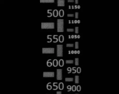
Great zoom range and high resolution
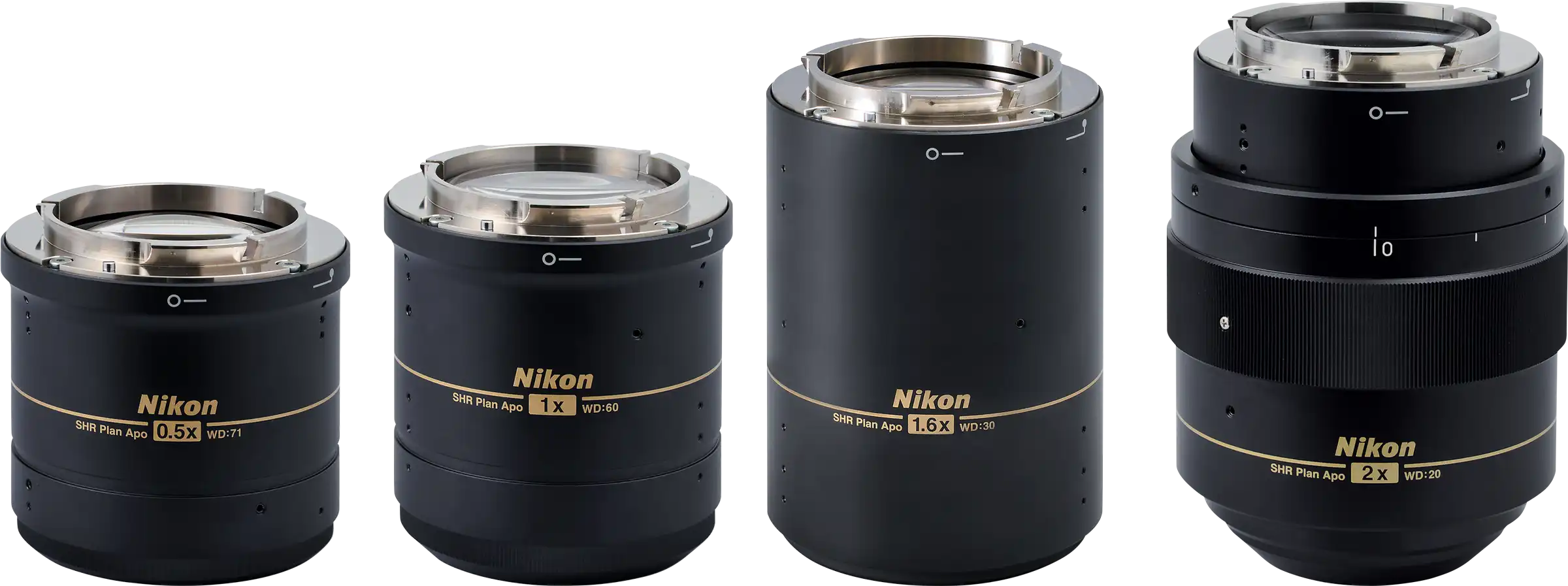
The SHR Plan Apo series objective lens offers a high resolution of 1100LP/mm (observed value, using SHR Plan Apo 2X at maximum zoom), delivering brilliant images with true-to-life colors.
Comparison of resolution and color aberration by resolution chart
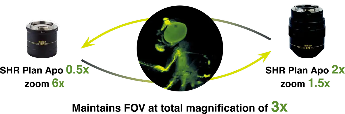
Auto Link Zoom (ALZ) supports seamless viewing at different scales
ALZ automatically adjusts the zoom factor to maintain the same field of view when switching objective lenses. This function enables seamless switching between whole organism imaging at low magnifications and detailed imaging at high magnifications.
Enhanced brightness and uniform illumination in low magnification range
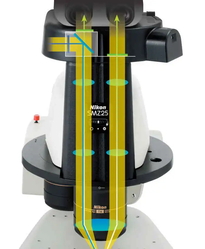
The SMZ25 series is the first stereo microscope in the world to use a fly-eye lens on an epi-fluorescence attachment. This innovative design ensures bright and uniform illumination even at low magnifications, resulting in uncompromised uniformity in brightness across a large field of view.
Conventional epi-fluorescence attachment New epi-fluorescence attachment
Poor illumination coverage Fly eye lens uniformly illuminates the entire field of view
Improved S/N ratio and crystal clear fluorescent images thanks to an improved optical system
Nikon's newly developed optical system offers a drastic improvement in S/N ratio even at high magnifications. This improved S/N ratio makes it possible to capture low fluorescence signals and light-sensitive events such as cell division, which is difficult using conventional stereo microscopes.
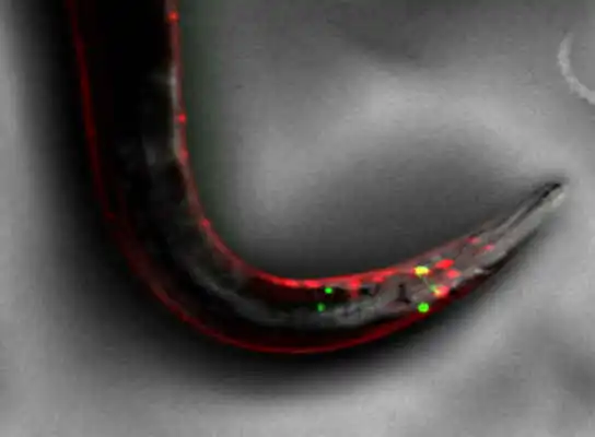
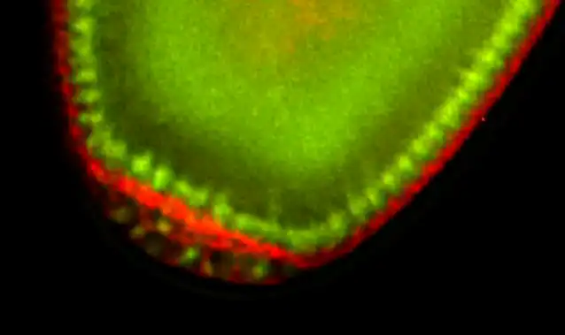
Single fluorescent neurons can be visualized in live C. elegans
Fluorescence and OCC images of a live C. elegans expressing GFP- and RFP-neurons
(using SHR Plan Apo 2X at zoom magnification of 3X with SMZ25)
Image courtesy of Julie C. Canman, Ph.D., Columbia University
Zoom body with significant improvements in optical performance
Nikon has succeeded in improving the signal and reducing noise in fluorescent images by using a short wavelength, high transmission lens. Combined with an innovative epi-fluorescence attachment, the SMZ18/SMZ25 is better able to detect emission signal than conventional fluorescent stereo microscopes.
Wide range of digital imaging capabilities
Easily obtain the information you need, such as Z drive position, zoom factor, objective lens, filter cube, and LED DIA brightness by using the Digital Sight series and NIS-Elements together with the microscope.
NIS-Elements Imaging Software
One software for all systems: NIS-Elements which is Nikon's flagship, cross-platform imaging software can also be used with Nikon's latest stereo microscope systems SMZ25 and SMZ18. NIS-Elements enables a wide range of advanced digital imaging capabilities.
Multichannel (multicolor)
Multiple fluorescent channels can be captured in conjunction with other imaging methods such as OCC or brightfield.
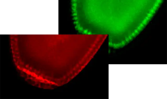
Images taken in individual colors Overlapping image with all colors
Individual cells resolved in a live drosophila embryo expressing GFP and mCherry (Using SHR Plan Apo 2X at zoom magnification of 8X with SMZ25)
Image courtesy of Max V. Staller, Ph.D., Clarissa Scholes, and Angela DePace, Ph.D., Harvard Medical School
Time-lapse
Easily setup a time-lapse imaging experiment with NIS-Elements.
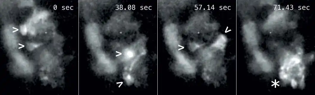
Calcium-imaging: Time-lapse imaging of GCaMP expressing neurons inside a live zebrafish shows individual neurons firing at different times (arrowheads). The last time-frame shows a whole cluster of neurons firing (asterisk).
(Using SHR Plan Apo 2X at zoom magnification of 9X with SMZ25 and camera head DS-Qi1)
Image courtesy of Joe Fetcho, Ph.D., Cornell University
Extended depth of focus (EDF)
Capture multiple high resolution images at different focal depths to create a single extended depth of focus image or quasi-3D image.
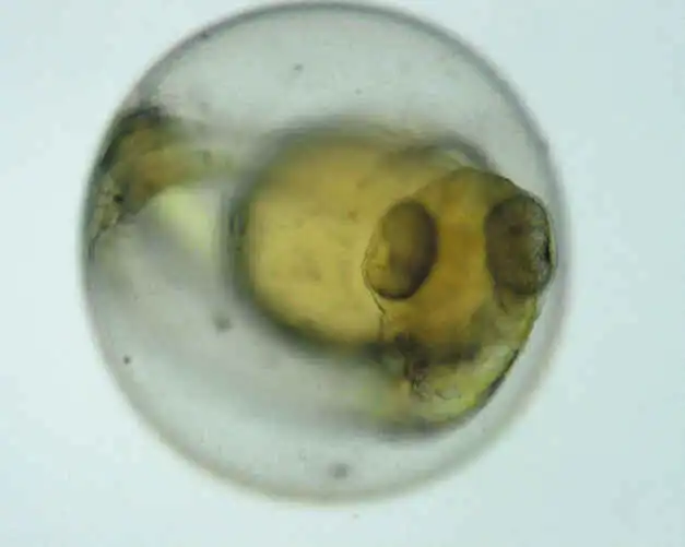

Select the in-focus area and produces one all-in-focus image Zebrafish embryo (Using SHR Plan Apo 2X at zoom magnification of 3.4X with SMZ25)
Image courtesy of Hisaya Kakinuma, Ph.D., Laboratory for Developmental Gene Regulation, Developmental Brain Science Group, RIKEN Brain Science Institute
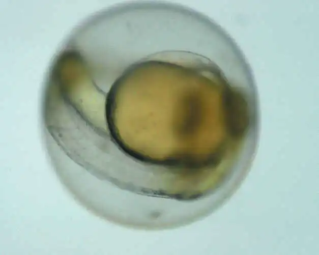
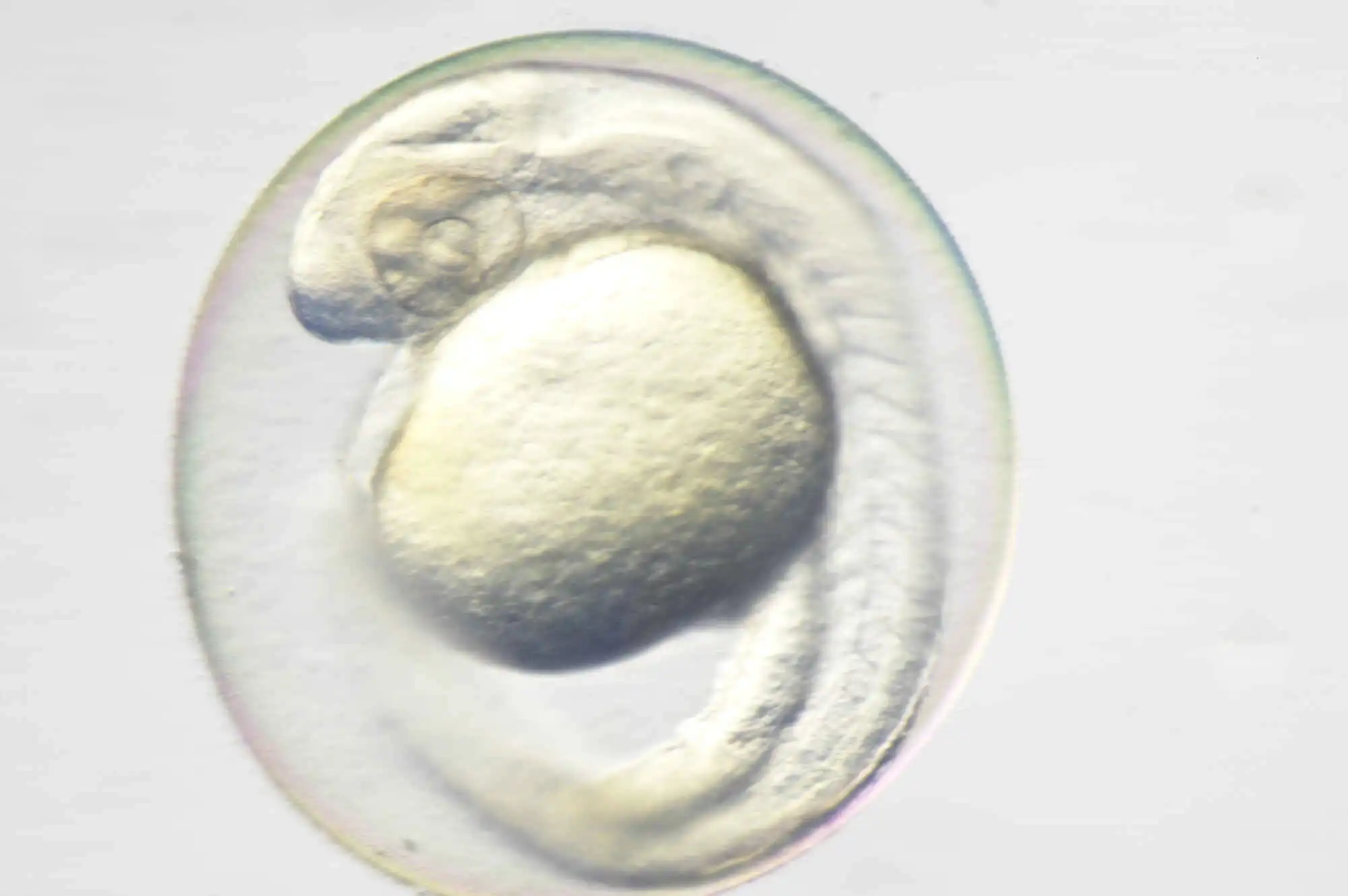
Easy-to-use OCC illumination
The new LED DIA Base with a built-in OCC illuminator generates minimal heat, consumes very little power and is long-lived. This illuminator can enhance the contrast of uneven surfaces, such as that of an embryo.
Conventional diascopic illumination OCC illuminator
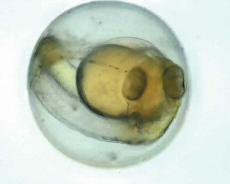
Zebrafish embryo (using SHR Plan Apo 1x at zoom magnification of 5x with SMZ18)
Image courtesy of Junichi Nakai, Ph.D. Saitama University Brain science Institute
The OCC illuminator can be controlled using a slide lever. Thanks to scales on the slide lever, the user can save and reproduce desired illumination levels. In addition, an OCC plate can be inserted into the illumination unit from the front and rear sides, so images with different shadow direction can be observed.
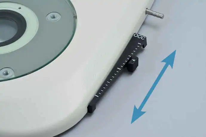
OCC illuminator
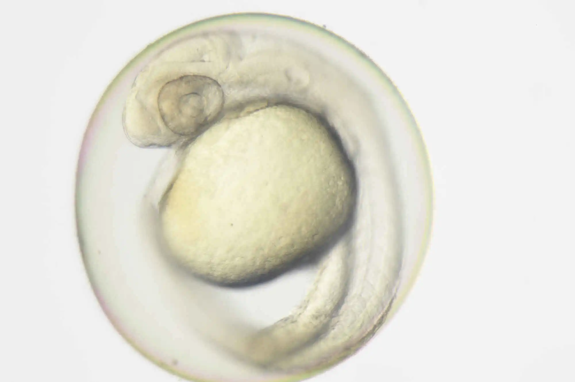
What is OCC illumination?
The acronym OCC stands for oblique coherent contrast (OCC), which is a form of oblique lighting method developed by Nikon. Compared to conventional diascopic illumination that illuminates directly from below, OCC illumination applies coherent light to samples in a diagonal direction, giving contrast to colorless and transparent sample structures.
Examples of OCC images


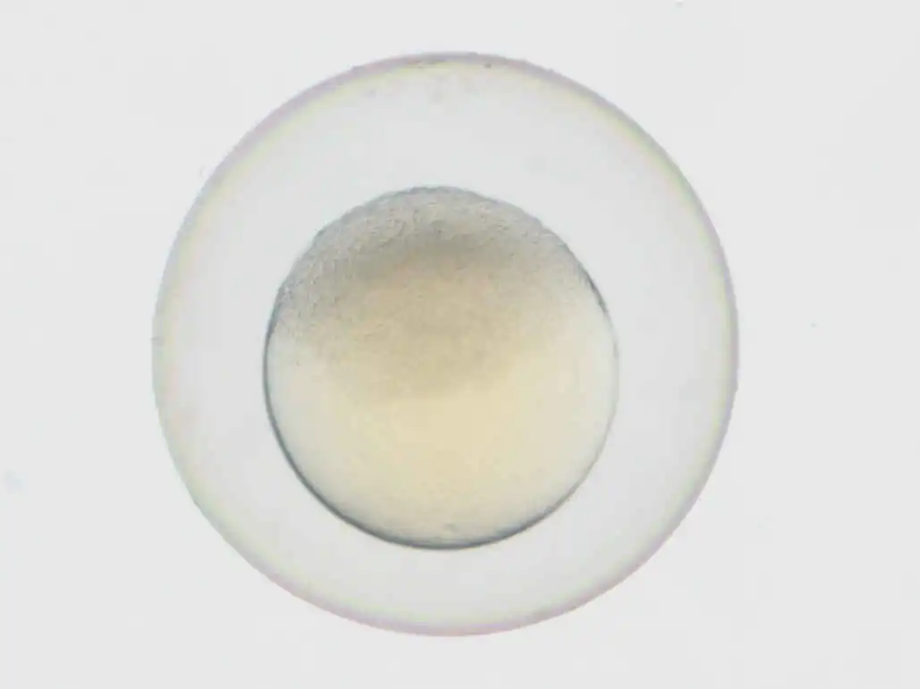
OCC illumination Standard transmitted illumination
C. elegans using SHR Plan Apo 1X, at zoom magnification of 13.5X, SMZ18 with P-DSF32
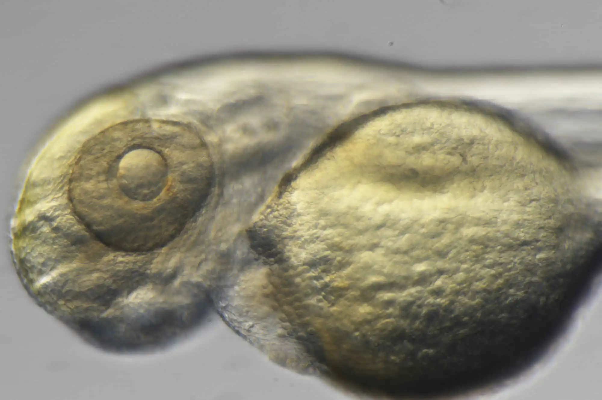
Fiber Diascopic Illumination
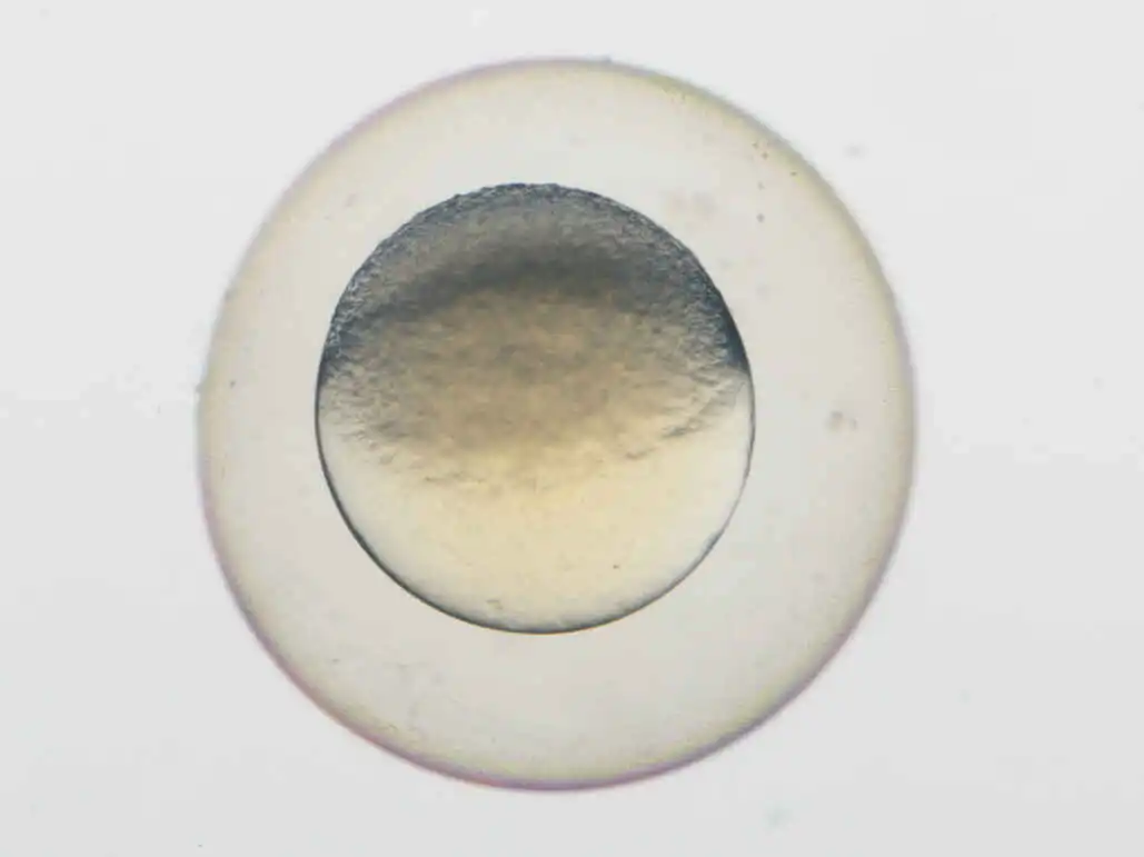
OCC illumination Standard transmitted illumination
1 day Zebrafish embryo using SHR Plan Apo 1X, at zoom magnification of 8X, SMZ18 with P-DSF32
Fiber Diascopic Illumination
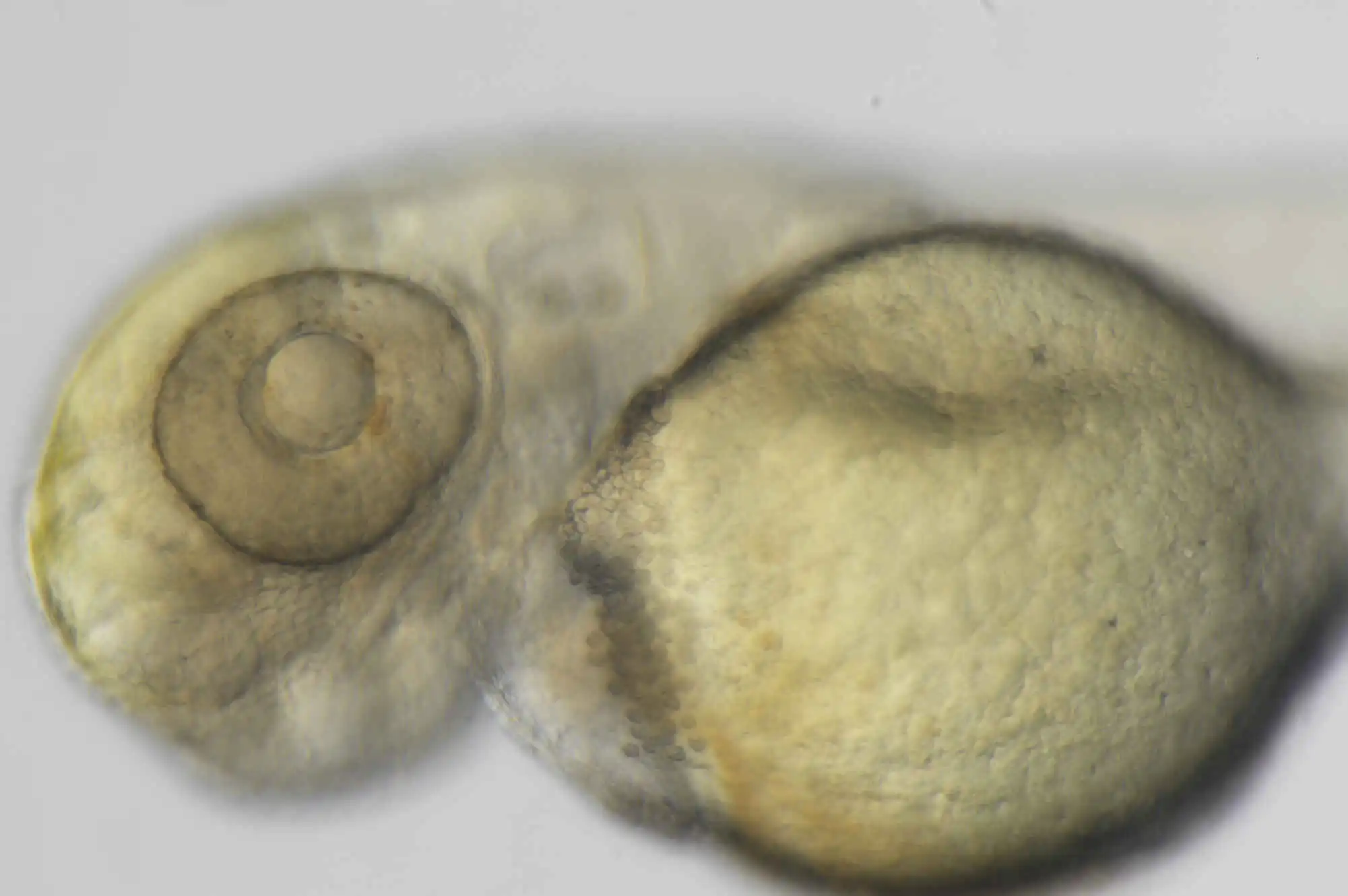
OCC illumination Standard transmitted illumination
2 days post hatching Zebrafish using SHR Plan Apo 1X, at zoom magnification of 13.5X, SMZ18 with P-DSF32
Fiber Diascopic Illumination
Image courtesy of:Hitoshi Okamoto, MD, PhD
Lab for Neural Circuit Dynamics of Decision Making RIKEN Center for Brain Science (CBS)
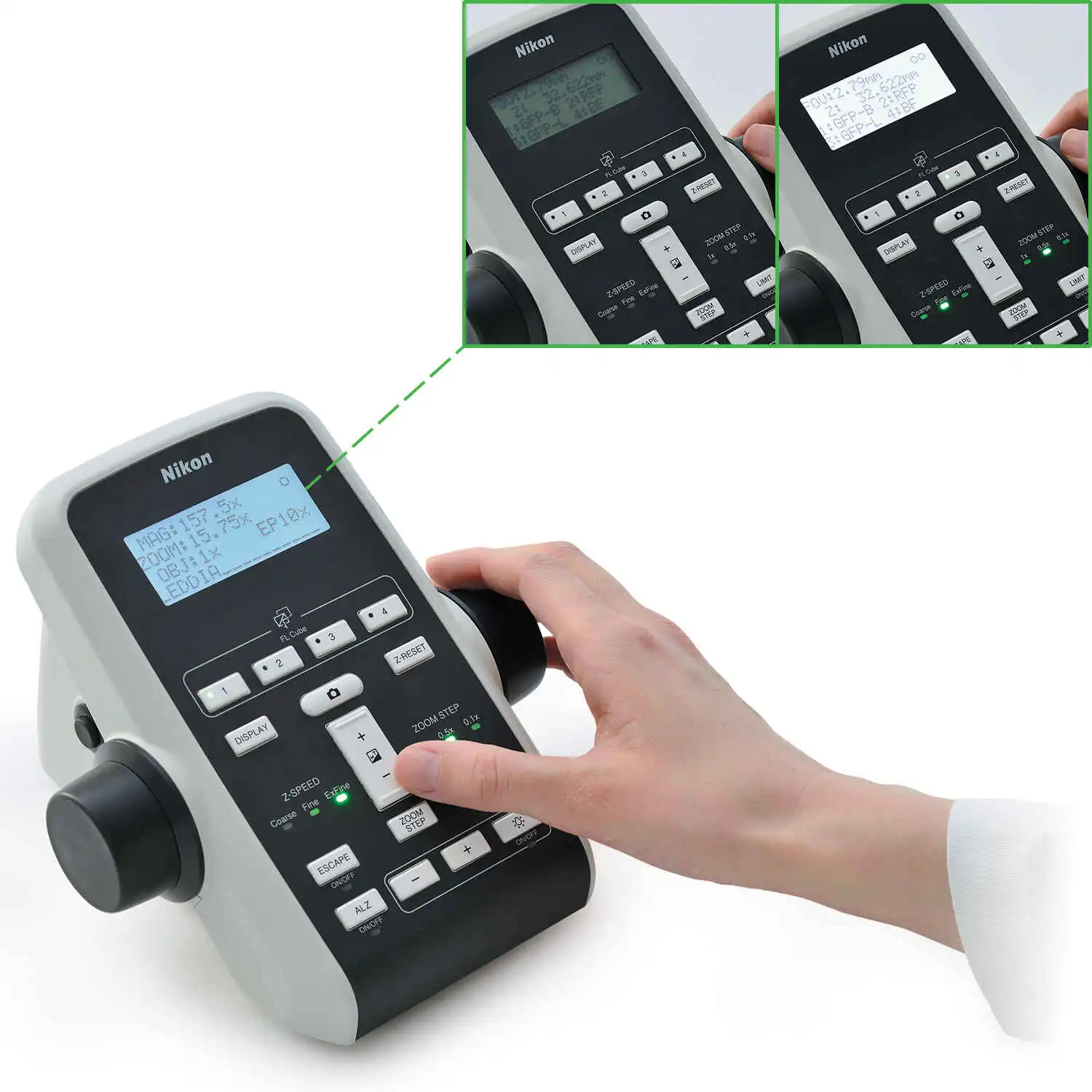
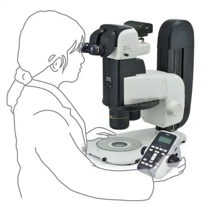
User-friendly remote controller
The remote controller provides easy access to zoom and focus controls and is designed for both right and left hand use. The remote controller contains an LCD monitor with an adjustable backlight which provides information regarding the zoom factor, objective lens, filter cube, and LED DIA brightness at a glance.
The remote controller contains an LCD monitor with an adjustable backlight which provides information regarding the zoom factor, objective lease, filter cube, and LED DIA brightness at a glance.
On-axis imaging for digital images
Easily switch between stereo position (stereoscopic view) and mono position (on-axis view) when using the P2-RNI2 Intelligent Nosepiece by simply sliding the objective lens.
Note: The affiliations and information are accurate as of the time of publication.


 Microsystem
 Endoscopysystem
 Energysystem
 EndoscopyConsumables
 +86-21-54286005
 Room 602, Building 1, No. 111 Luxiang Road (Greenland Park Plaza), Baoshan District, Shanghai, China
  English
 中文
中文
 SMZ25 SMZ18_Brochure_EN


 中文
中文 English