Olympus GF-UCT260
EVIS LUCERA Ultrasound Gastrovideoscope
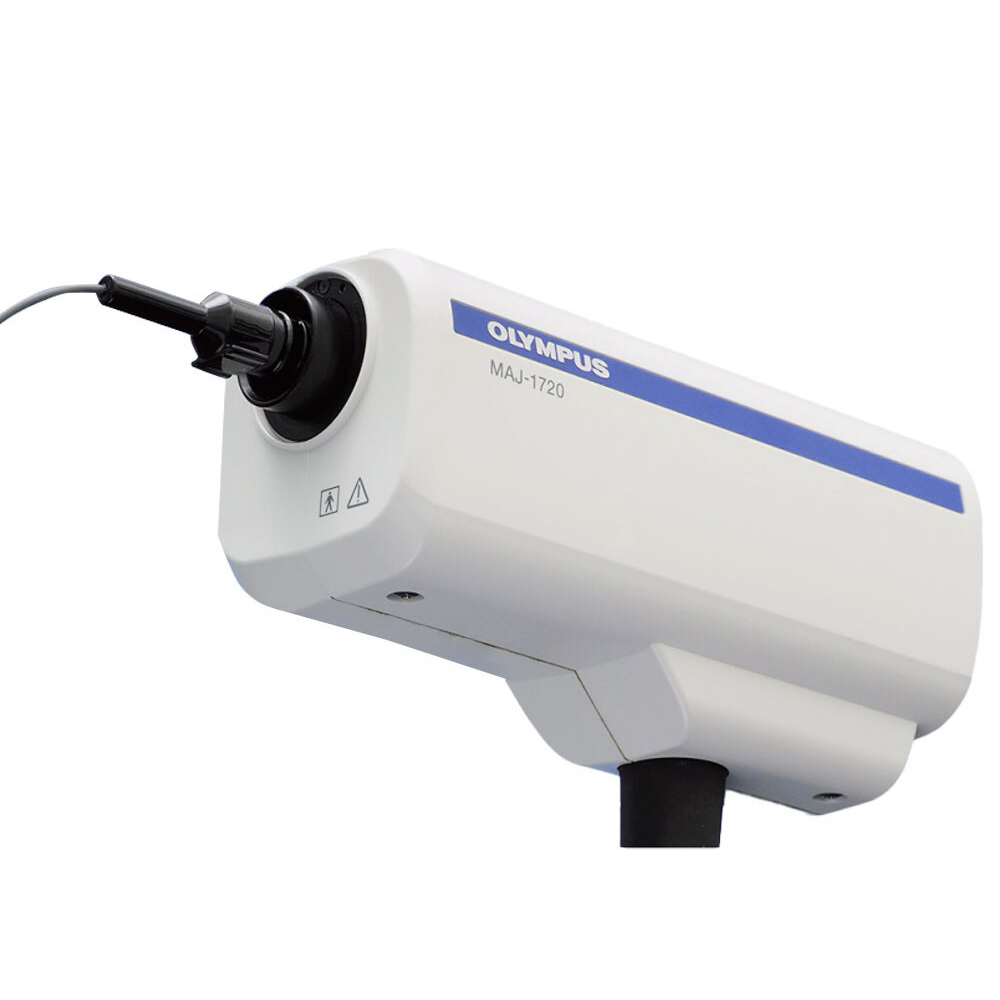
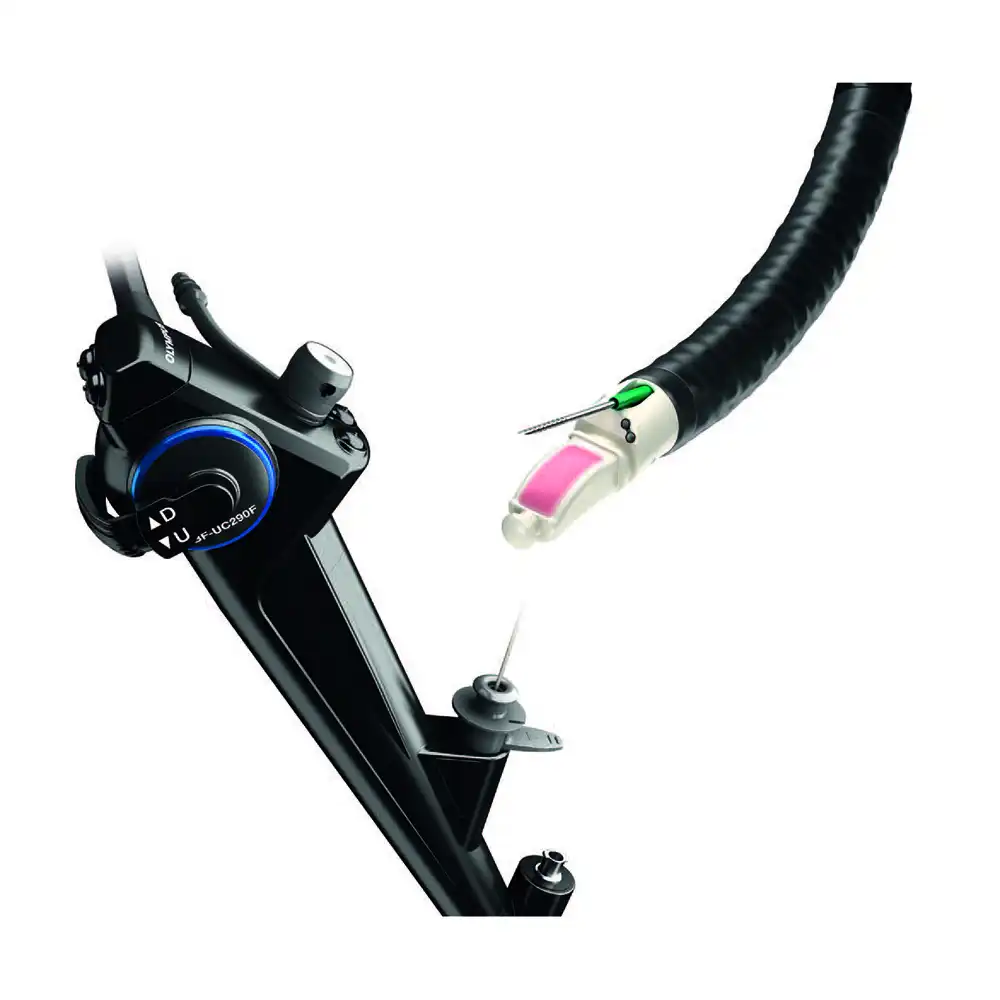
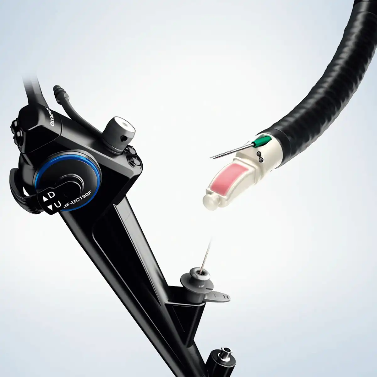
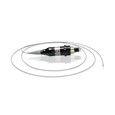
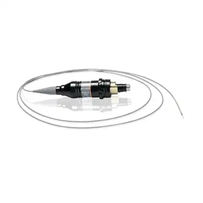
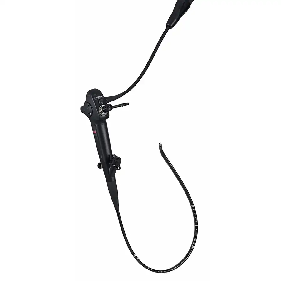
The new GF-UCT260 EVIS LUCERA ultrasound gastrovideoscope provides improved imaging depth and a redesigned forceps elevator to facilitate accurate needle placement. A single detachable cable connects this versatile scope with a choice of ultrasound centres, providing flexibility for echoendoscopy procedures. The latest GF-UCT260 EVIS LUCERA ultrasound gastrovideoscope delivers high quality endoscopic ultrasound images with greater imaging depth. The innovative forceps elevator design facilitates finely controlled needle handling during endoscopic ultrasound treatment, whilst the large 3.7 mm instrument channel accommodates bigger needles for more advanced fine needle aspiration procedures. A single detachable cable connects this versatile scope with a choice of ultrasound centres, providing the ultimate in flexibility for echoendoscopy procedures.

Outstanding compatibility
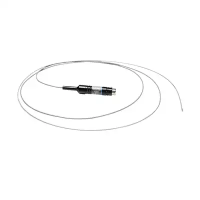
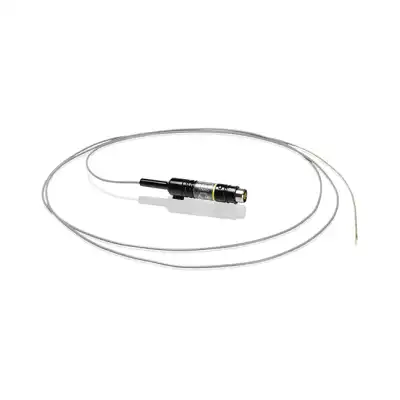
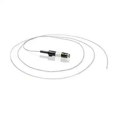
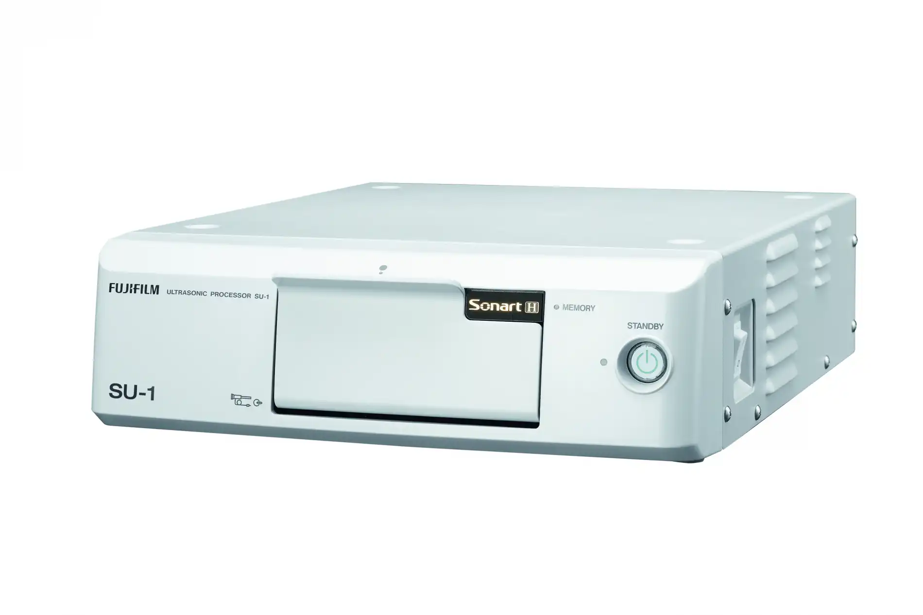
A single detachable cable (MAJ-1597) enables connection to a range of compatible Olympus and Hitachi Aloka ultrasound centres, providing the ultimate in platform flexibility.
Excellent device handling
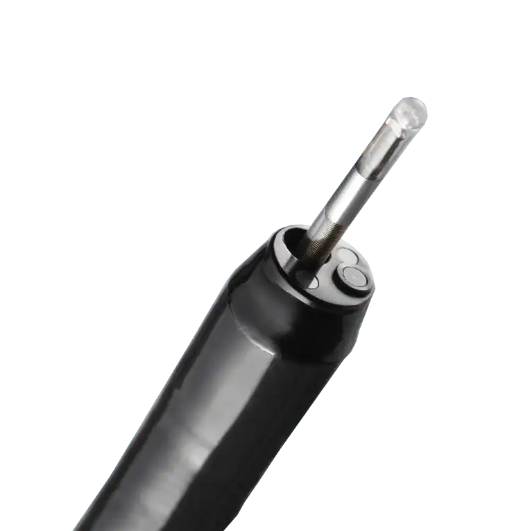
An innovative new design of forceps elevator enables fine control and handling of devices inserted through the 3.7 mm diameter instrument channel for reliable fine needle aspiration.
Platform flexibility
A single detachable cable enables connection to a range of compatible Olympus and Hitachi Aloka ultrasound centres.
Redesigned forceps elevator
The redesigned forceps elevator provides improved fine control and handling of devices inserted through the 3.7 mm diameter instrument channel.
Large 3.7 mm diameter channel
The exceptional 3.7 mm diameter working channel accommodates larger aspiration needles of over 22G, enabling more advanced FNA techniques.


 Microsystem
 Endoscopysystem
 Energysystem
 EndoscopyConsumables
 +86-21-54286005
 Room 602, Building 1, No. 111 Luxiang Road (Greenland Park Plaza), Baoshan District, Shanghai, China
  English
 中文
中文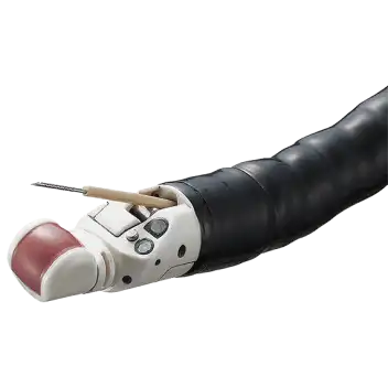
 GF-UCT260_Brochure_EN


 中文
中文 English