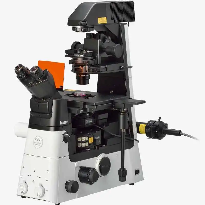
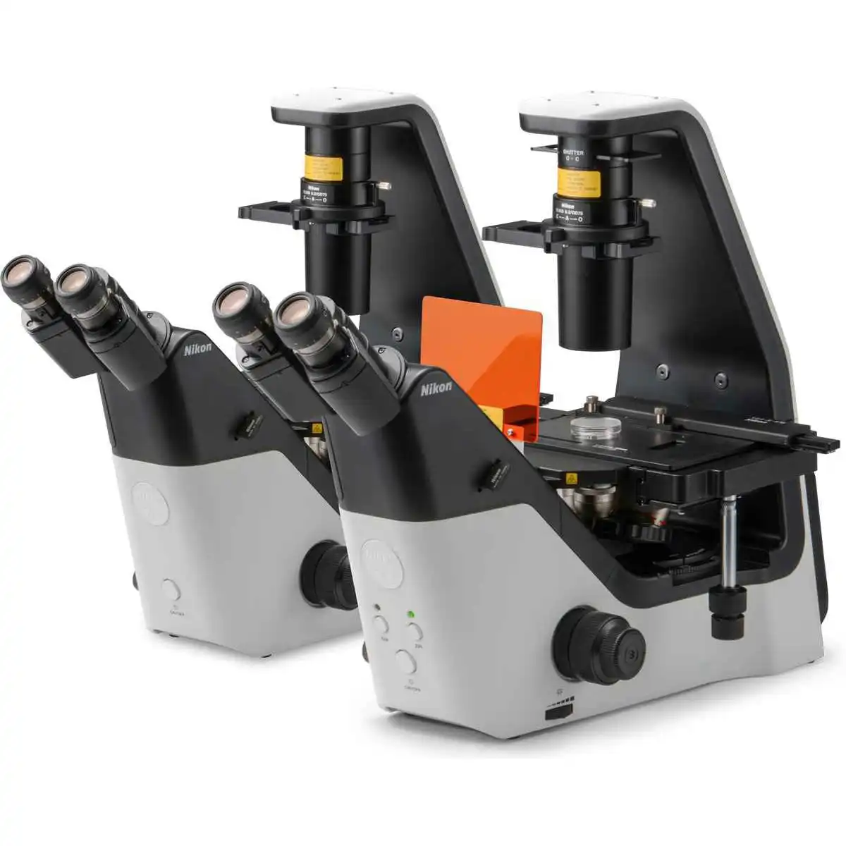
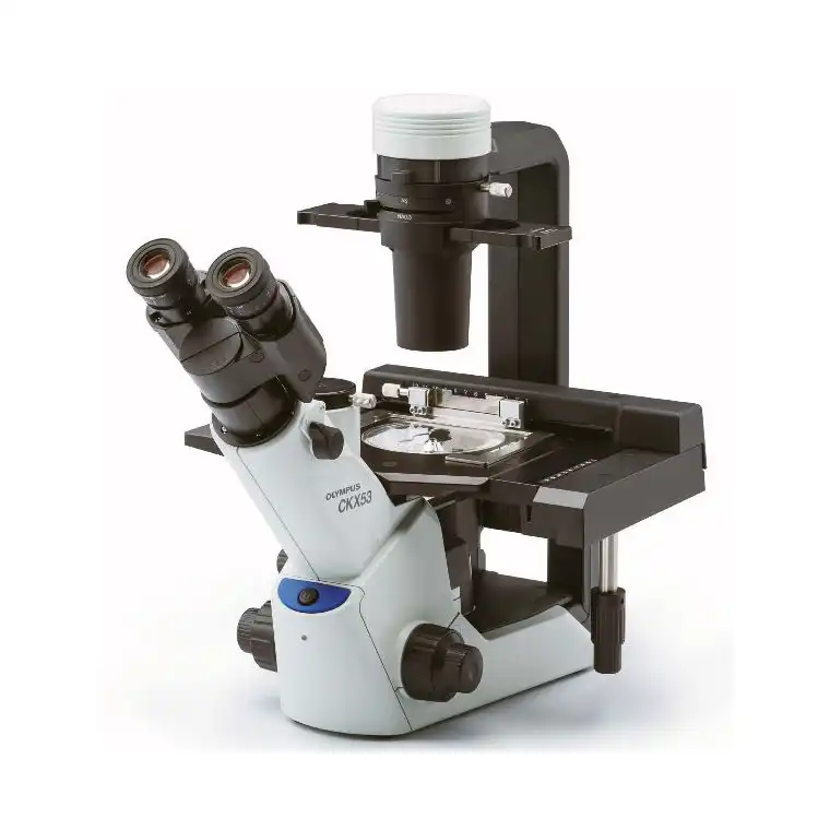
NIKON ECLIPSE Ti2-A
Research Inverted Microscope
Leading platform for advanced imaging.
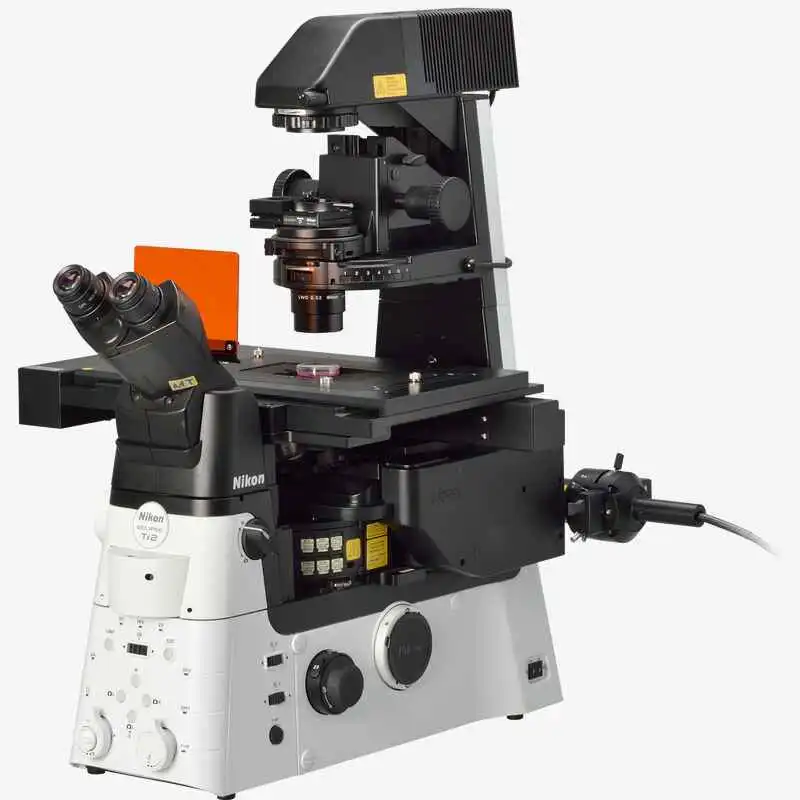
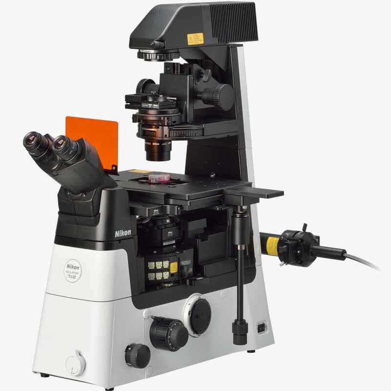
The ECLIPSE Ti2-A inverted microscope delivers an unparalleled 25mm field of view (FOV) that revolutionizes the way you see. With this incredible FOV, the Ti2-A maximizes the sensor area of large-format CMOS cameras without making compromises, and significantly improves data throughput. The Ti2-A's exceptionally stable, drift-free platform is designed to meet the demands of super-resolution imaging while its unique hardware-triggering capabilities enhance even the most challenging, high-speed imaging applications. Furthermore, the Ti2-A's unique, intelligent functions guide users through imaging workflows by gathering data from internal sensors, eliminating the possibility of user errors. In addition, the status of each sensor is automatically recorded during acquisition, providing quality control for imaging experiments and enhancing data reproducibility.
In combination with Nikon's powerful acquisition and analysis software, NIS-Elements, the Ti2-A is a total innovation in imaging.
Key Features
Groundbreaking FOV
As research trends evolve towards large-scale, systems-level approaches, there is an increasing demand for faster data acquisition and higher throughput capabilities. Development of large-format camera sensors and improvements in the data processing capabilities of PCs have facilitated such research trends. The Ti2-A, with its unprecedented 25mm field of view, provides the next level of scalability, enabling researchers to truly maximize the utility of large-format detectors and future-proof their core imaging platform as camera technologies continue to develop at a rapid pace.
Bright illumination over a wide area
High-power LEDs deliver bright illumination across the Ti2-A's large field of view, ensuring clear, consistent results from demanding applications such as high-magnification DIC. Incorporation of a fly-eye lens design provides uniform illumination from edge to edge for quantitative high-speed imaging and seamless tiling of images in stitching applications.

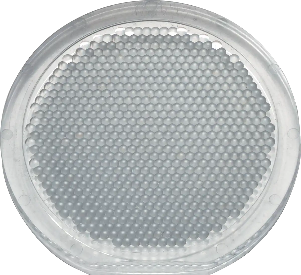
High-power LED illuminator Built-in fly-eye lens
A compact epi-fluorescence illuminator designed for large FOV imaging is equipped with a quartz fly-eye lens and provides high transmittance across a broad spectrum, including UV. Large diameter fluorescence filters with hard coatings deliver large FOV images with a high signal-to-noise ratio.

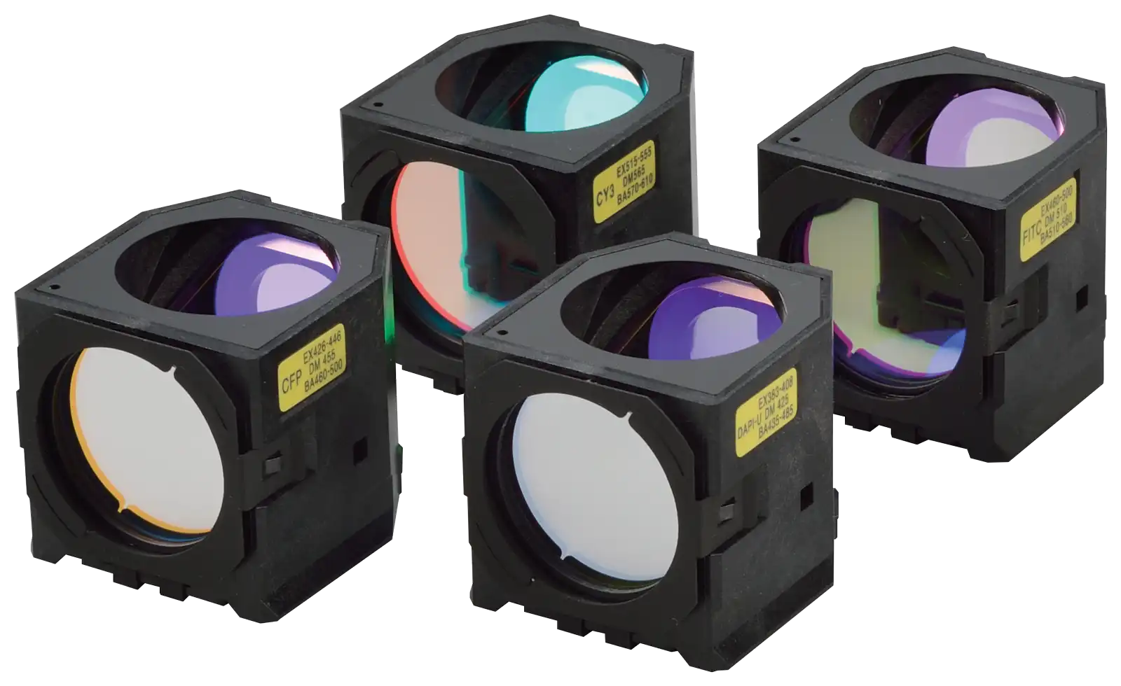
Large FOV epi-fl illuminator Large diameter fluorescence filter cubes
Large diameter observation optics
The diameter of the observation light path has been enlarged in order to achieve a field number of 25 at the imaging port. The resulting large FOV is capable of capturing approximately double the area of conventional optics, enabling users to gain maximum performance from large-format sensors such as CMOS detectors.

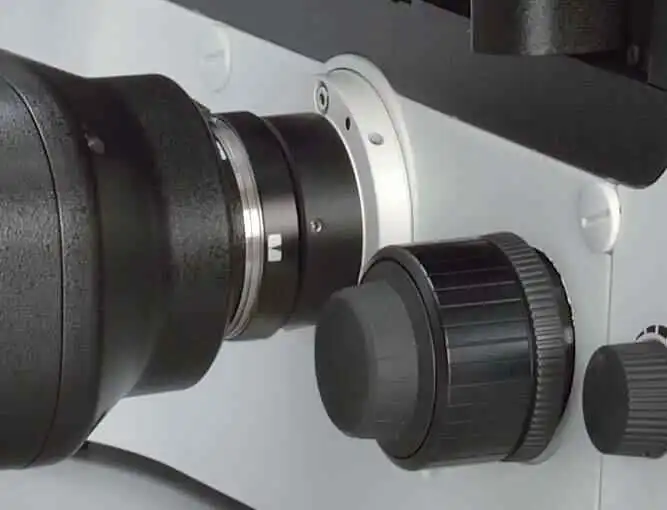
Enlarged tube lens Imaging port with large 25 field number
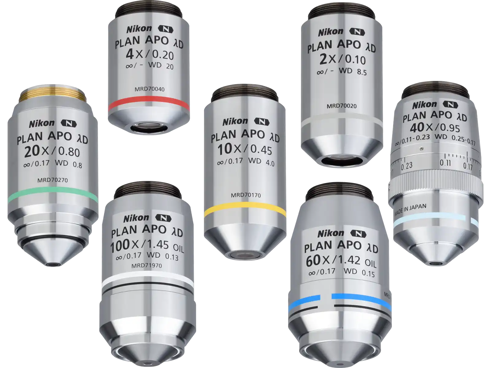
Objectives for large FOV imaging
Objectives with superior image flatness ensure high quality images from edge to edge. Utilizing the maximum potential of the OFN25 objective significantly accelerates data collection.
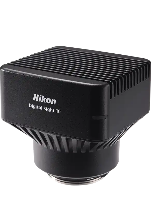
Cameras for large-volume data acquisition
Nikon's FX-format F-mount cameras Digital Sight 50M and Digital Sight 10 are equipped with CMOS image sensors optimized for research use, originally developed for professional D-SLR cameras. This allows high-speed and high-sensitivity live-cell imaging, enabling the best use to be made of the Ti2's large FOV.


Unsurpassed Nikon optics
Nikon's high-precision CFI60 infinity optics, designed for use with a variety of sophisticated observation methods, are highly regarded by researchers for their superb optical performance and solid reliability.
Apodized phase contrast
Nikon's unique apodized phase contrast objectives with selective amplitude filters dramatically increase contrast and reduce halo artifacts to provide detailed high-definition images.

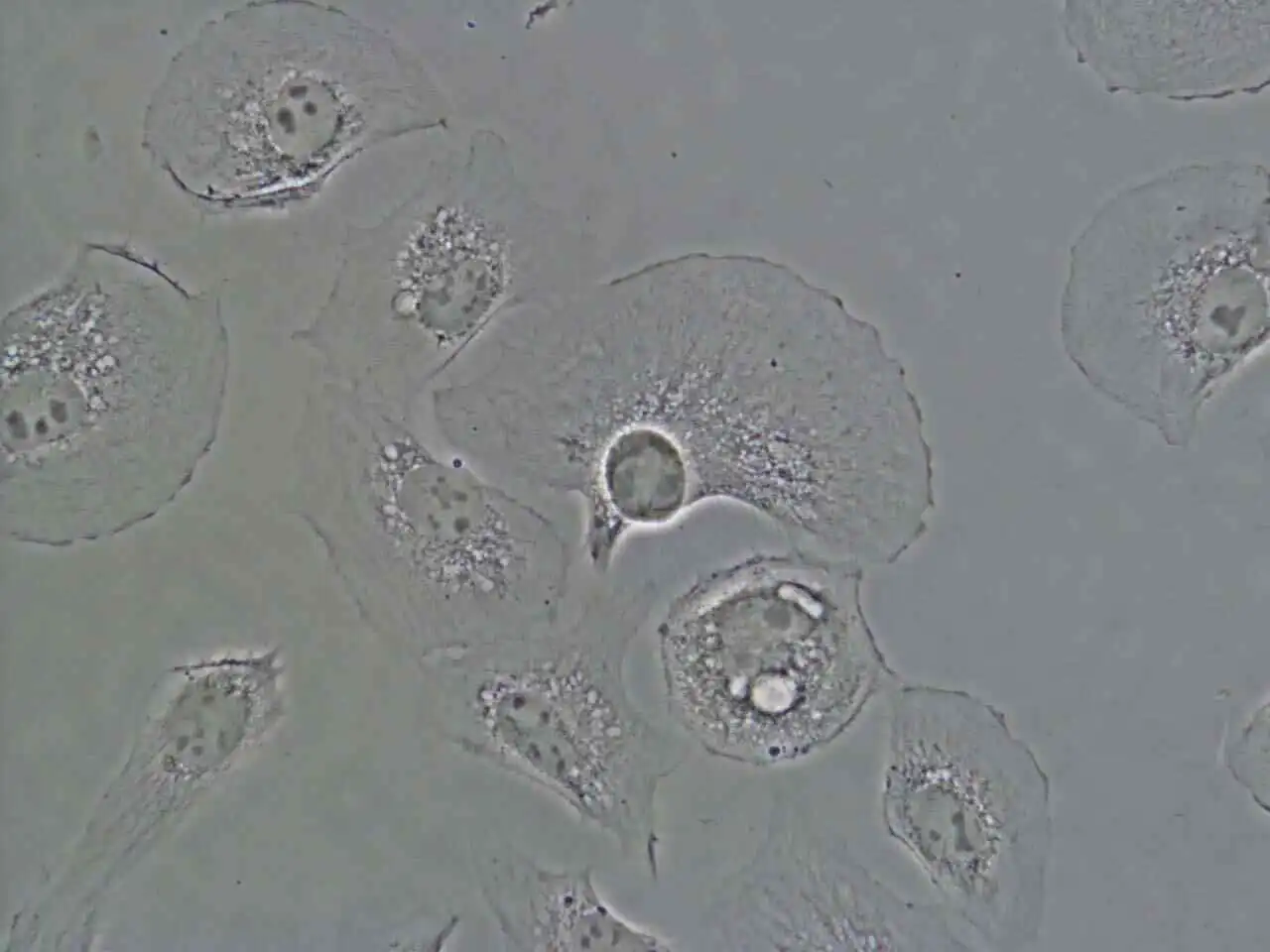
Apodizedphase plate is incorporated in APC objectives BSC-1 cells captured with CFI S Plan Fluor ELWD ADM 40XC objective
DIC (Differential Interference Contrast)
Nikon's highly-regarded DIC optics provide uniformly clear and detailed images with high resolution and contrast throughout the magnification range. DIC prisms are individually tailored for each objective lens to provide the highest-quality DIC images for every sample.

DIC prisms matched to individual objectives are mounted in the nosepiece
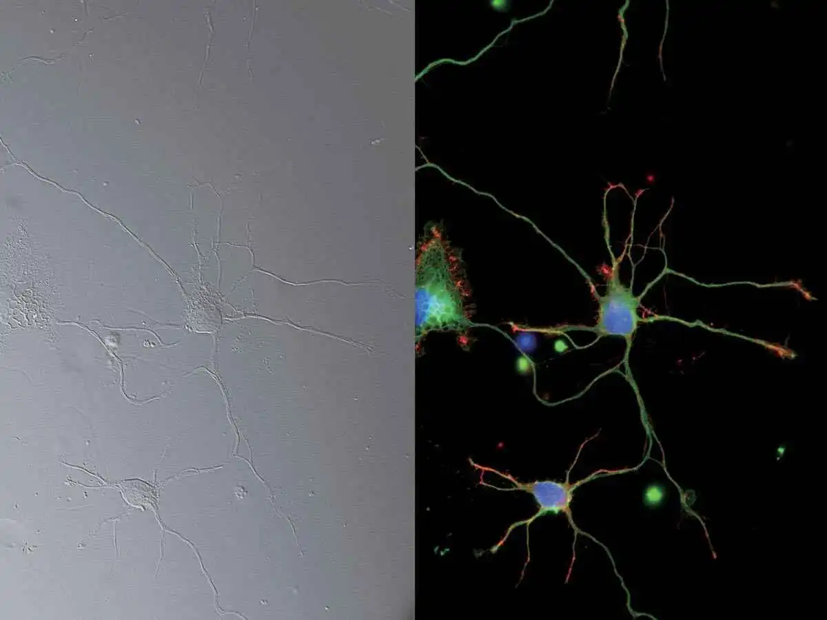
DIC and epi-fluorescence images:
25mm FOV image of neurons (DAPI, Alexa Fluor® 488, Rhodamine-Phalloidin), captured with CFI Plan Apochromat Lambda 60XC objective and DS-Qi2 camera
Photo courtesy of Josh Rappoport, Nikon Imaging Center, Northwestern Univ.;
Sample courtesy of S. Kemal, B. Wang, and R. Vassar, Northwestern Univ.
NAMC (Nikon Advanced Modulation Contrast)
This is a plastic-compatible, high-contrast imaging technique for unstained, transparent samples such as oocytes. NAMC provides pseudo-three-dimensional images with a shadow-cast appearance. The direction of contrast can be easily adjusted for each sample.


NAMC objective lenses contain rotatable modulators NAMC image:Mouse embryos, captured with CFI S Plan Fluor ELWD NAMC 20XC objective
Epi-fluorescence
The Lambda series objectives, utilizing Nikon's proprietary Nano Crystal Coat technology, are perfect for demanding, low-signal, multi-channel fluorescence imaging that requires high transmission and aberration correction over a wide wavelength range. Combined with new fluorescence filter cubes that offer improved fluorescence detection and stray light countermeasures such as the Noise Terminator, the Lambda series objectives demonstrate their power in weak signal observations such as single-molecule imaging and even luminescence-based applications.

Luminescence image:
HeLa cells expressing BRET-based calcium indicator protein, Nano-lantern (Ca2+).
Sample courtesy of Prof. Takeharu Nagai, The Institute of Scientific and Industrial Research, Osaka University
Continuous display of microscope status
A collection of built-in sensors detects and relays status information for a variety of components in the microscope. All of the status information is recorded in the metadata when you acquire images with a computer, so you can easily recall acquisition conditions and/or check for configuration errors.
In addition, a built-in internal camera allows users to view the back aperture, facilitating confirmation of phase ring alignment and extinction cross in DIC. It also provides a laser-safe method for aligning lasers for applications such as TIRF.
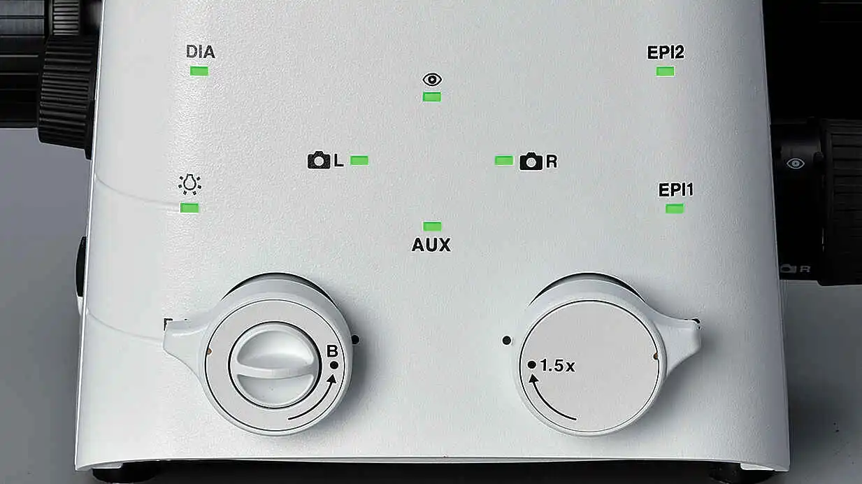
Status Lights
Guidance for operational procedures
The Ti2's Assist Guide function provides interactive step-by-step guidance for microscope operation. The Assist Guide can be viewed on a tablet or PC, and integrates real time data from built-in sensors and an internal camera. The Assist Guide is designed to help users through alignment procedures for both experiment setup and troubleshooting.
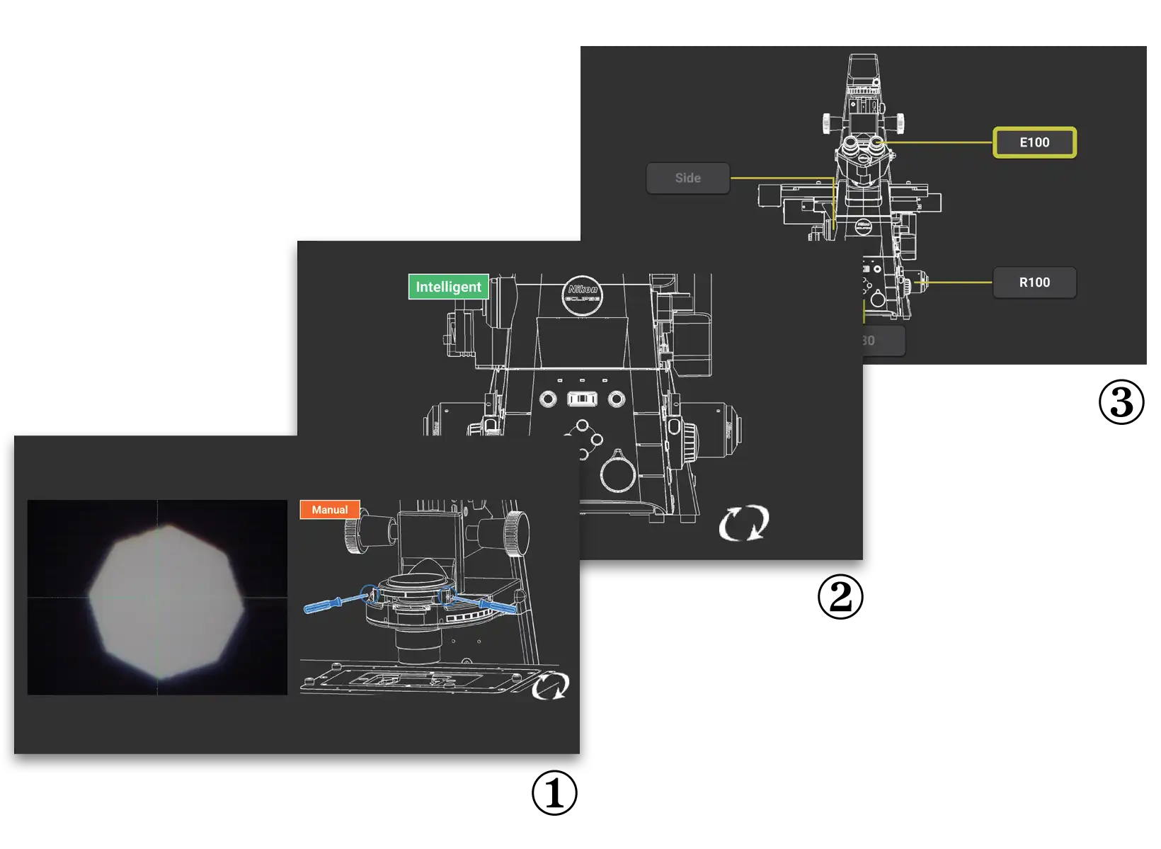
① Move the field diaphragm image to the center of the field view
② Remove the Bertrand lens from the optical path
③ Select an observation port
Automatically detect errors
The Check Mode allows users to easily confirm, on either a tablet or PC that all the correct microscope components are in place for their chosen observation method. This capability eliminates time and effort normally required for troubleshooting when the desired observation method is not achieved. This functionality is particularly advantageous when multiple users are involved, each with the potential to make unexpected changes to the microscope settings. Custom check procedures can also be pre-programmed.
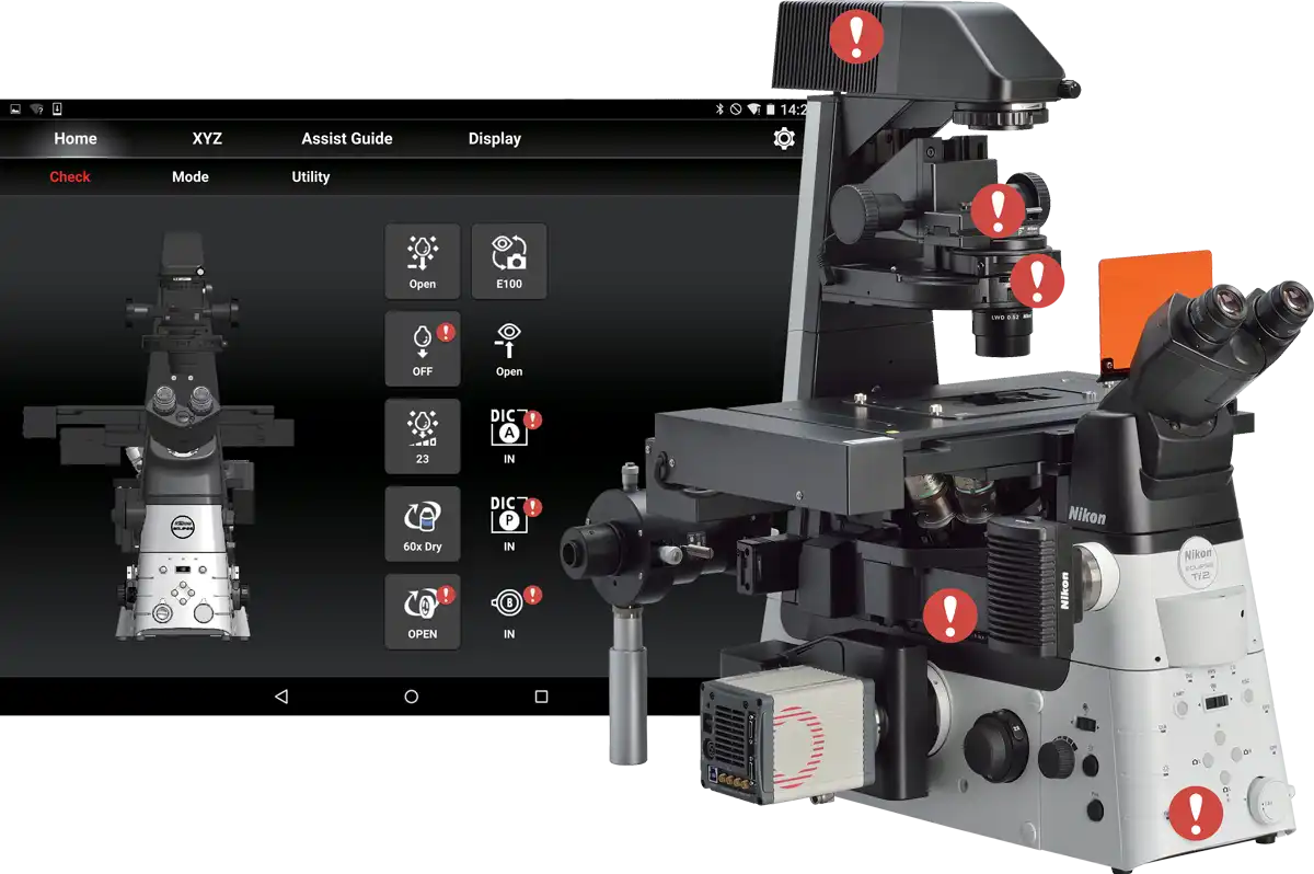
Displays misaligned components
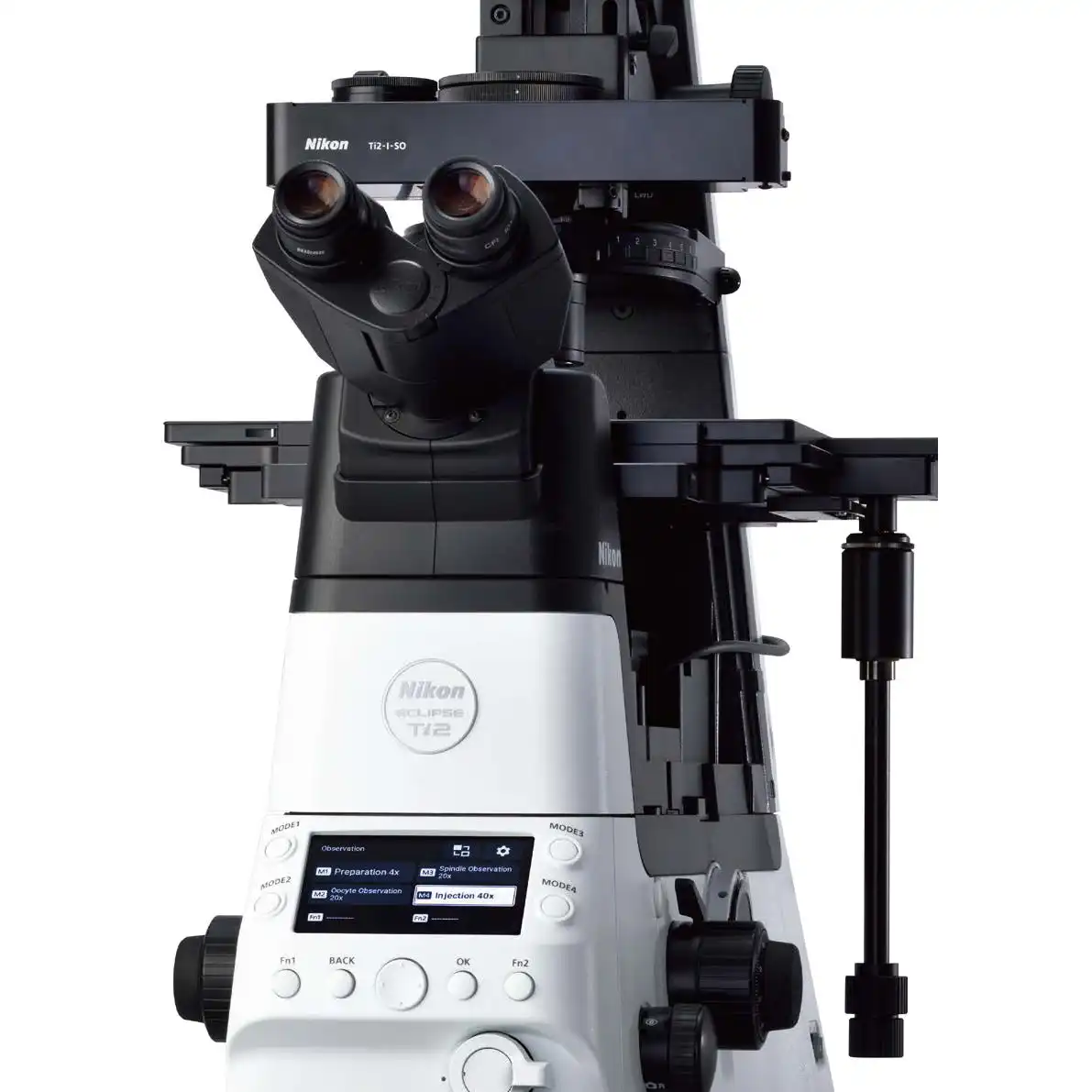
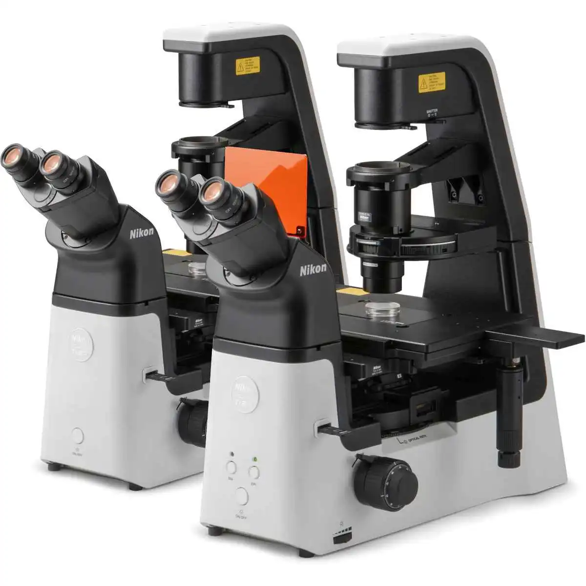
Intuitive operation
The Ti2-A has been completely redesigned, from the overall body design to the selection and placement of every button and switch, for the ultimate in user experience. The controls are easy to use even in the dark, where the majority of imaging experiments are conducted. The Ti2-A provides an intuitive and effortless user interface so researchers can focus on the data and not on microscope control.
Thoughtfully designed layout for microscope control
The placement of all of the buttons and switches are based on the type of illumination they control. Buttons that control diascopicobservation are positioned on the left side of the microscope and those that control epi-fluorescence observation are on the right side. Buttons that control common operations are on the front panel. This use of zoning provides an easy-to-remember layout, a desirable feature when operating the microscope in a dark room.
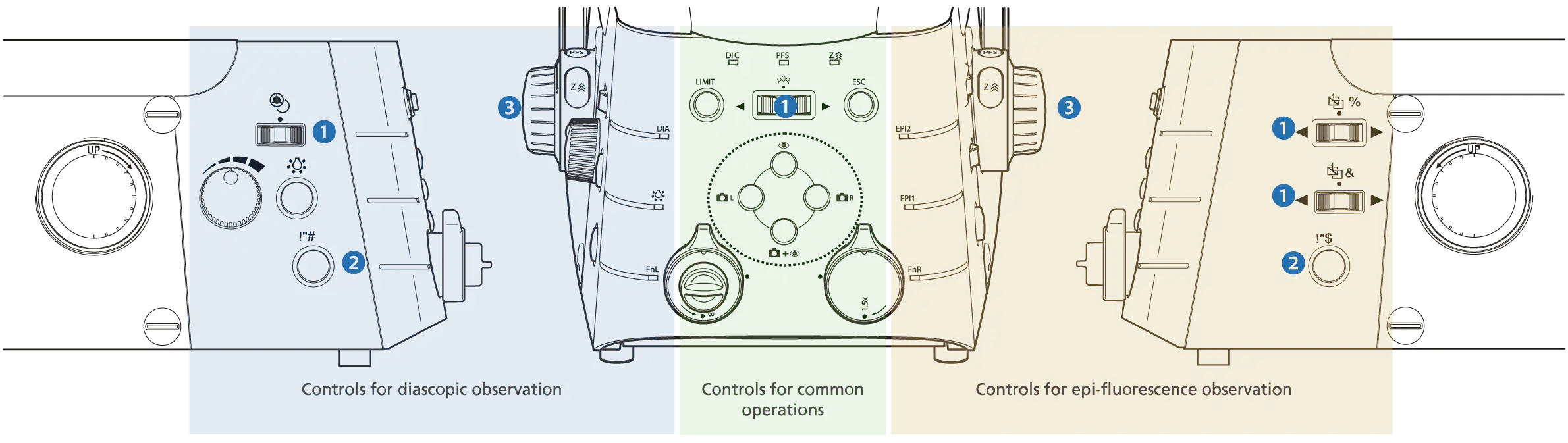
② Programmable Function button (Ti2-E/A)
Conveniently located Function buttons allow customization of the user interface. Users can select from more than 100 functions, including control of motorized devices such as shutters and even signal output to external devices via the I/O port for triggered acquisition. Mode functions, which enable instant changing of observation methods by storing the settings of each motorized device, can also be assigned to these buttons.
| Main body |
ECLIPSE Ti2-A |
| Optical system |
Infinity-corrected CFI60 |
| Field number*2 |
22 with C-mount, 25 with F-mount |
| Intermediate Magnification switching |
Manual switching of 1.0X/1.5X (exchangeable from 1.5X to 2.0X) |
| Output port |
4 Manual positions
Eyepiece 100%, left 100%, right 100%, option (to eyepiece
20%/left 80% or eyepiece 20%/right 80%) |
| Can add ports by use of back port unit and/or choice of tube base unit*3 |
| Focusing unit |
Manual drive, Coarse/fine focusing knob, 10 mm stroke |
| Stage up |
Available*4 |
| Binocular tube |
Binocular S tube TC-T-TS (field number 22), Ergonomic ER tube TC-T-ER (field number 22) |
| Assist eyepiece tube base unit (Ti2-T-BA) |
Assist camera (field number 22), Status detection |
| Pillar for transmitted illumination (Ti2-D-PD) |
Condenser vertical stroke: 66 mm, Backward tilting up to 25 degrees, With field diagram and refocusing mechanism 2 filter slot positions (4 filter position option is also available with Filter Slider for transmitted illumination (Ti2-D-SF)) |
| LED Lamphouse for dia illumination (Ti2-D-LHLED) |
High power LED |
| High Color Rendering LED Lamphouse (C-LL) |
High color rendering LED |
| Precentered Lamphouse (D-LH/LC) |
100W halogen bulb (pre-centered) |
| Intelligent condenser turret (Ti2-C-TC-I) |
7 manual positions (ø37 mm x4, ø39 mm x3), Status detection, LWD/ELWD/CLWD/NAMC condenser lenses are supported |
| Condenser turret (TC-C-TC) |
7 manual positions (ø37 mm x4, ø39 mm x3), LWD/ELWD/CLWD/NAMC condenser lenses are supported |
| ELWD-S condenser turret (TE-C) |
4 manual positions, With ELWD condenser lens (NA0.3/O.D.=65) |
| HNA condenser slider (Ti2-C-SCH) |
2 manual positions (ø37 mm x1, ø39 mm x1), HNA dry lens/HNA oil lens are supported |
| Condenser lens |
LWD (O.D.=30 mm, NA=0.52), ELWD (O.D.=75 mm, NA=0.29), CLWD (O.D.=13 mm, NA=0.72), HNA dry (O.D.=5 mm, NA=0.85), HNA oil (O.D.=2 mm, NA=1.4), NAMC (O.D.=44 mm, NA=0.4) |
| Stage (TC-S-SR, TC-S-SRF) |
Stroke X: ±57 mm, Stroke Y: ±36.5 mm, Adjustable stroke range (3 levels) with adjusting pin, Long/middle/short handle options available |
| Gliding stage (TC-S-GS) |
Stroke ø20 mm |
| Intelligent DIC sextuple nosepiece (Ti2-N-ND-I) |
6 manual positions, Status detection, Water-resistant*5- |
| Sextuple nosepiece (Ti2-N-N), DIC sextuple nosepiece (Ti2-N-ND) |
6 manual positions, Water-resistant*5 |
| Intelligent epi filter turret (Ti2-F-FLT-I) |
6 manual positions, Manual shutter, Status detection*6 |
| Motorized shutter (Ni-SH-E)*7 |
12ms to open/close |
| EPI-FL module (TI2-LA-FL-3) |
Supports fiber illuminator; includes 2-position filter slider and aperture diaphragm |
| Field stop slider |
Circular (TI2-F-FSC), rectangular (TI2-F-FSR), square (TI2-F-FSS) aperture options |
| Controller, display device |
Tablet |
| Operating environmental |
Temperature: 0℃+40℃, Humidity: 60% RH max. (at +40℃, no condensation), Indoor use only |
Motorized accessories have a status detection function
*1 Motorized model with a bottom port
*2 Limitations apply based on objective and filter cube choice, stage-up configuration, and illumination module, etc.
*3 Tube base units with a port cannot be used with Ti2-A
*4 Stage up kit is required. Please contact Nikon.
*5 This product is not waterproof. Water-resistant properties are only guaranteed when all specified conditions below are satisfied.
*6 Status detection cannot be used when attached to the Ti2-U
*7 Ni-SH-CON Controller for Motorized Shutter is required for use with the Ti2-A/Ti2-U
Conditions to Ensure Water-resistant Function of the Nosepiece
- Properly attach either of the following to the nosepiece:
- Objective lens mount: Objective lens or Objective lens mounting hole cap
- DIC slider mount: DIC slider cap or DIC slider
- Properly attach either the supplied water collection tray or the optional Ti2-N-PT nosepiece protection tray.
- Exercise caution to ensure that water discharged from the drainage tube does not come into contact with the main unit.
For the safe operation of this product, please follow the instructions in the user manual for proper installation and use.


 Microsystem
 Endoscopysystem
 Energysystem
 EndoscopyConsumables
 +86-21-54286005
 Room 602, Building 1, No. 111 Luxiang Road (Greenland Park Plaza), Baoshan District, Shanghai, China
  English
 中文
中文
 ECLIPSE Ti2_Brochure


 中文
中文 English