 EndoscopyConsumables
 English
 中文
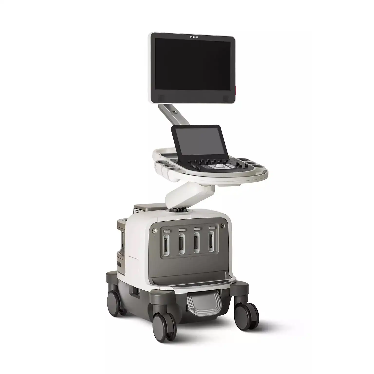
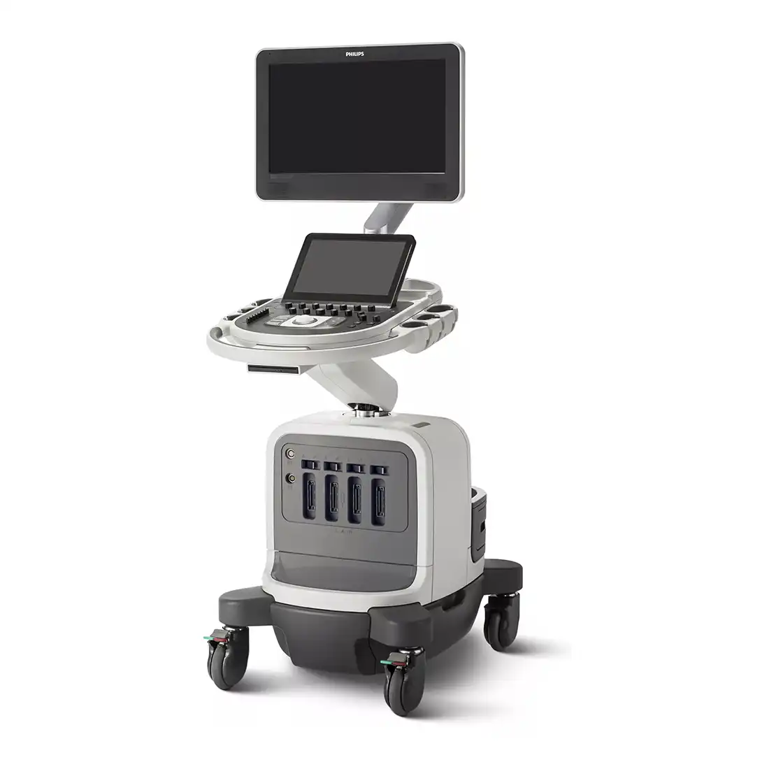
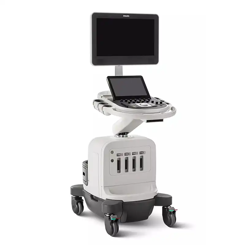
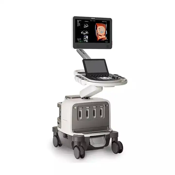
中文PHILIPS EPIQ Elite Ultrasound system
PHILIPS EPIQ Elite Ultrasound system
Phone:+86-21-54286005
Resources
 EPIQ Elite_Brochure_EN


 Microsystem
 Endoscopysystem
 Energysystem
 +86-21-54286005
 info@tenmed.net
 Room 602, Building 1, No. 111 Luxiang Road (Greenland Park Plaza), Baoshan District, Shanghai, China
PHILIPS EPIQ Elite Ultrasound system
Phone:+86-21-54286005
EPIQ Elite_Brochure_EN

EPIQ Elite Elevate provides high-quality imaging and tailored clinical information to help clinicians deliver timely, confident answers to more patients worldwide. With advanced intelligence and an exceptional level of performance, EPIQ Elite meets the demands of today’s most challenging practices.
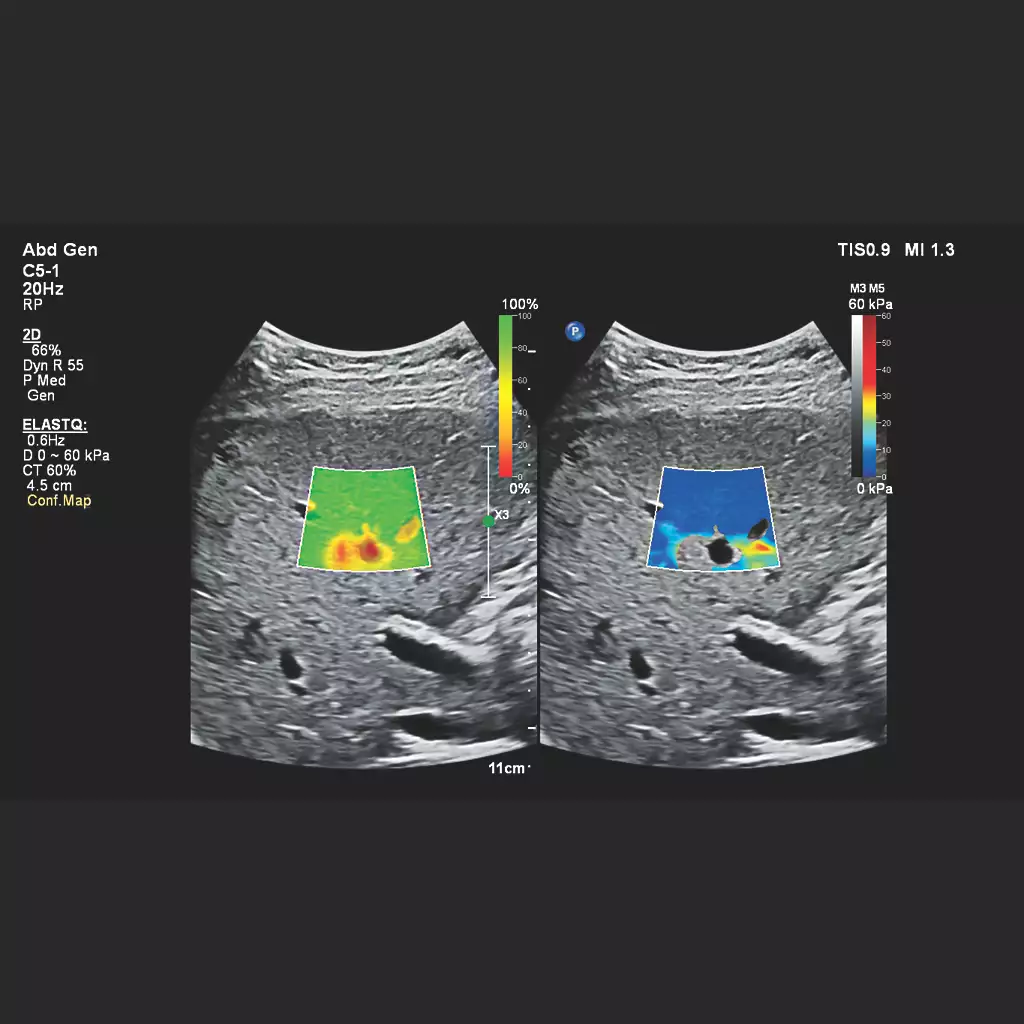
Auto ElastQ
Perform automated liver elastography with Auto ElastQ, and experience our next generation of liver health assessment. Auto ElastQ is designed to simplify user workflow with real-time, quantitative shear wave measurements.
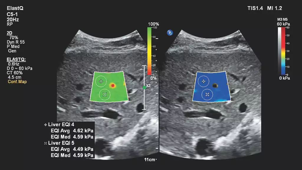
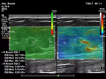
Elastography
The EPIQ Elite platform supports both strain and shear wave imaging methods of elastography. Highly sensitive strain imaging can be used to rapidly assess relative tissue stiffness values across a variety of applications. ElastQ Imaging methods of shear wave elastography use a unique pulsing scheme to generate and detect the propagation speed of shear waves, providing a quantitative display and measurement of tissue stiffness. ElastQ Imaging also provides a confidence map display to assist you in obtaining measurements from areas with the highest shear wave quality.
CEUS high frame rate linear & Auto Scan
See a 67%*** increase in CEUS frame rate and a 76%*** increase field of view with the eL18-4 transducer when thyroid scanning. CEUS Auto Scan improves image uniformity and sensitivity
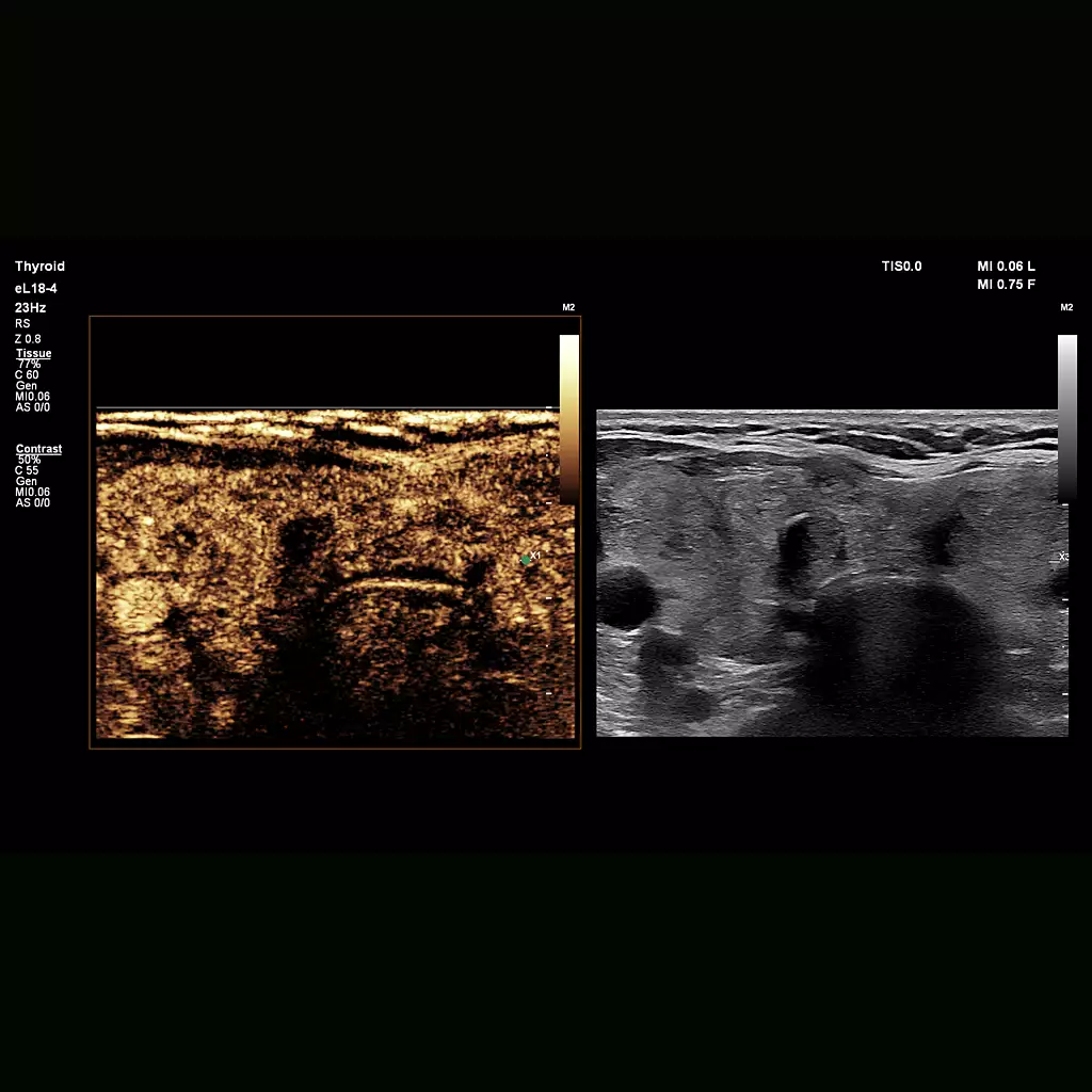
Contrast-enhanced ultrasound (CEUS)
CEUS can transform the role of ultrasound in the liver, allowing the study of the enhancement patterns of suspicious liver lesions in real time, as well as provide an alternative non-ionizing approach to the assessment of vesicoureteral reflux in pediatric patients.
_EN.webp)
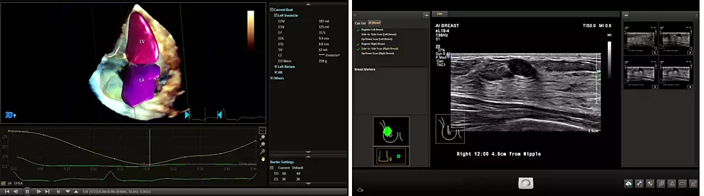
AutoStrain LV with automated EF and mid-layer strain
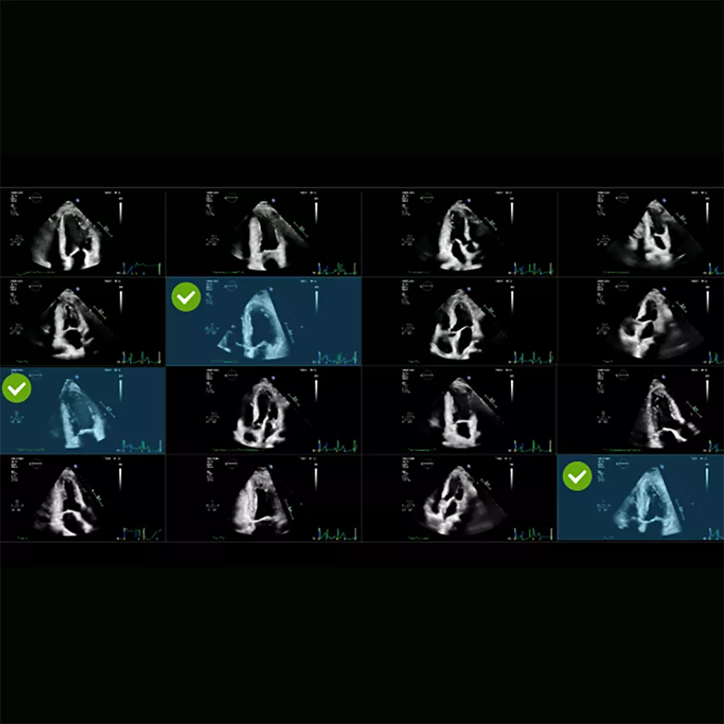
Advances to AutoStrain feature fast, reproducible results as part of a comprehensive LV assessment within the same application, improving workflow and saving time. Smart View Select works in the background and uses AI to automatically select the optimum images for 2D LV assessment.
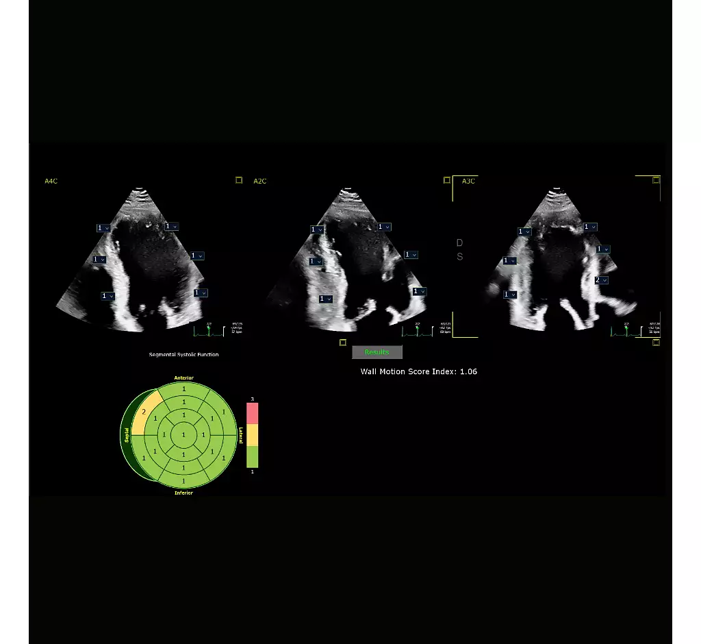
Auto Segmental Wall Motion Scoring
Provides automated evaluation of wall motion in a standard 17-segment bullseye display to aid objective LV wall assessment. With Auto SWMS, you can achieve greater reproducibility and efficiency in your workflows.
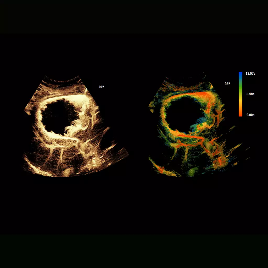
Super Resolution MVI & Time of Arrival
Philips Super Resolution MVI CEUS is an improved version of Philips legacy Contrast MVI which takes advantage of an innovative super-resolution processing approach and advanced motion compensation techniques to obtain a 200% improvement in spatial resolution. The Time of arrival map offers representation of the relative time at which bubbles first enter points of interest in the current imaging plane.
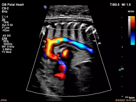
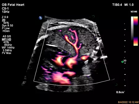
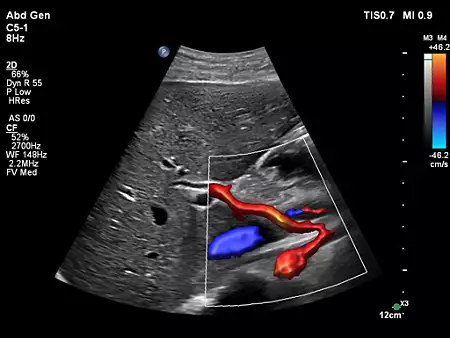
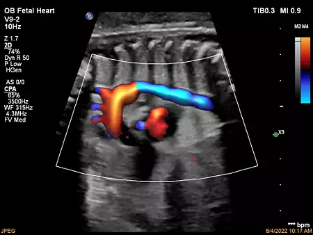
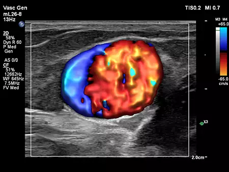
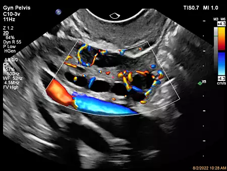
Flow Viewer
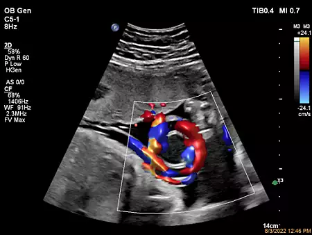
Flow Viewer is a Philips color visualization enhancement for vasculature and fetal heart architecture. Flow Viewer provides a 3D-like rendering of flow imaging data to better visualize the cardiac and vascular architecture and enhance the aesthetic appeal of all color imaging modes Available in all color imaging modes (CFM, CPA, CPAd, MFI, MFI HD).
.webp)
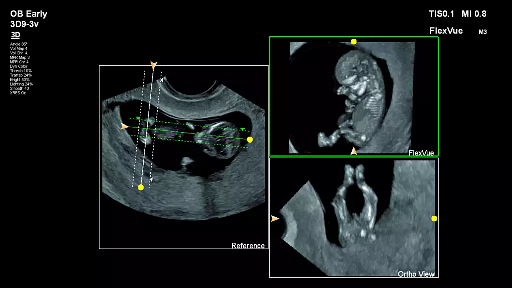
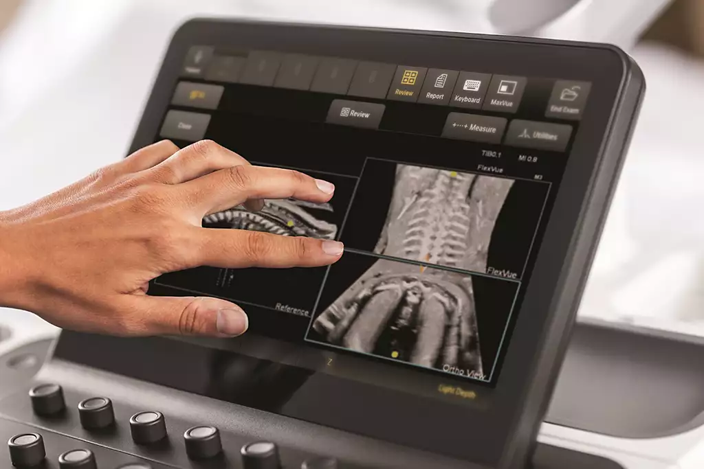
FlexVue with Orthogonal View
Easy-to-use tools designed to extract challenging anatomical planes from 3D data sets. This advanced feature offers exceptional flexibility in plane acquisition, complemented by a comprehensive measurement package for precise quantification.
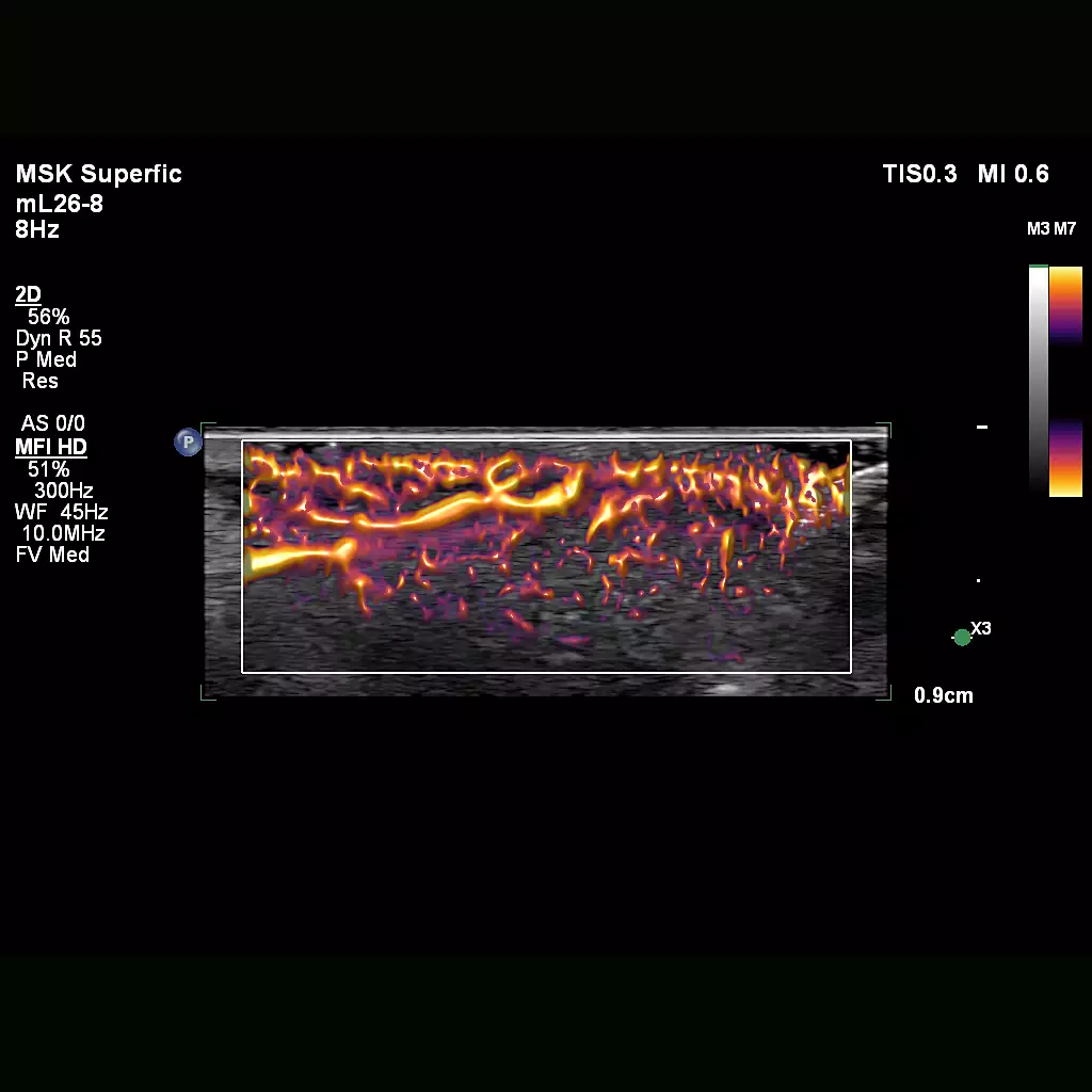
MFI HD
Designed to detect slow and weak blood flow anatomy in tissue. This proprietary approach overcomes many of the barriers associated with conventional methods to detect small vessel blood flow with high resolution and minimal artifacts. MicroFlow Imaging maintains high frame rate and 2D image quality while applying advanced artifact reduction techniques to reveal small vessel anatomy
Next Gen Auto Scan
Philips Next Gen Auto Scan improves image uniformity, adaptively adjusting image brightness at every pixel and reducing the need for user adjustment while also improving transducer plunkability. Next Gen Auto Scan reduces button pushes by up to 54% with pixel-by-pixel real-time optimization.
Collaboration Live
Extend your team without expanding it. Collaboration Live is a communication platform that facilitates communication between a compatible ultrasound system and a remote user. With simultaneous Multi-party communication up to six users can quickly and securely talk, text, screen share and video stream directly from the ultrasound system for access to multiple clinical resources at a distance.

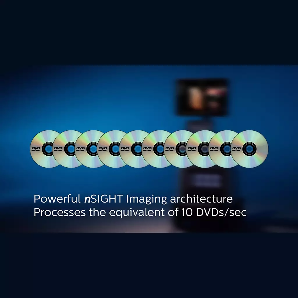
nSIGHT Imaging
Far surpasses conventional ultrasound performance to reach new levels of definition and clarity. Incorporating a custom multi-stage precision beamformer along with massive parallel processing, this proprietary architecture captures an enormous amount of acoustic data from each transmit operation and performs digital beam reconstruction along with mathematically optimized focal processing. This creates extraordinary real-time images with exceptional frame rate, uniformity and penetration.
XRES Pro, the next-generation image processing
At real-time frame rates, XRES Pro uses multi-parametric precision filters that subdivide image elements, analyze this data and then apply advanced algorithms to sharpen borders and interfaces and provide superb tissue conspicuity. XRES Pro also offers enhanced assessment of plaque morphology. XRES Pro allows you full adjustability to match the level of enhancement to clinical imaging requirements for elevated diagnostic confidence with virtually all patients.
MicroFlow Imaging
Designed to detect slow and weak blood flow anatomy in tissue. This proprietary approach overcomes many of the barriers associated with conventional methods to detect small vessel blood flow with high resolution and minimal artifacts. MicroFlow Imaging maintains high frame rate and 2D image quality while applying advanced artifact reduction techniques to reveal small vessel anatomy
PureWave & xMatrix transducer technology
The power of PureWave for exceptional imaging even on technically difficult patients. PureWave crystal technology represents the biggest breakthrough in piezoelectric transducer material in 40 years. The pure, uniform crystals of PureWave have virtually perfect uniformity for greater bandwidth and twice the efficiency of conventional ceramic materials.
xMatrix transducers, powerful and versatile
No other premium ultrasound solution can run xMatrix, the comprehensive suite of the world’s most innovative ultrasound transducers. Achieve ultra-thin 2D slices. Use Live xPlane imaging to create two full-resolution planes simultaneously, allowing you to capture twice as much clinical information in the same amount of time. Acquire near-isovoxel resolution to reveal images from any plane within the volume.
24" HD MAX display
This new immersive 24” display monitor offers the ultimate ultrasound visualization experience, with an ultra-wide color gamut of 10-bit color depth that uses billions of colors for accurate color reproduction. In addition, it provides high-contrast dynamic range and enhanced black levels for subtle delineation of grayscale values. HD MAX features superb off-angle viewing for visualization of clinical images throughout the scanning room.
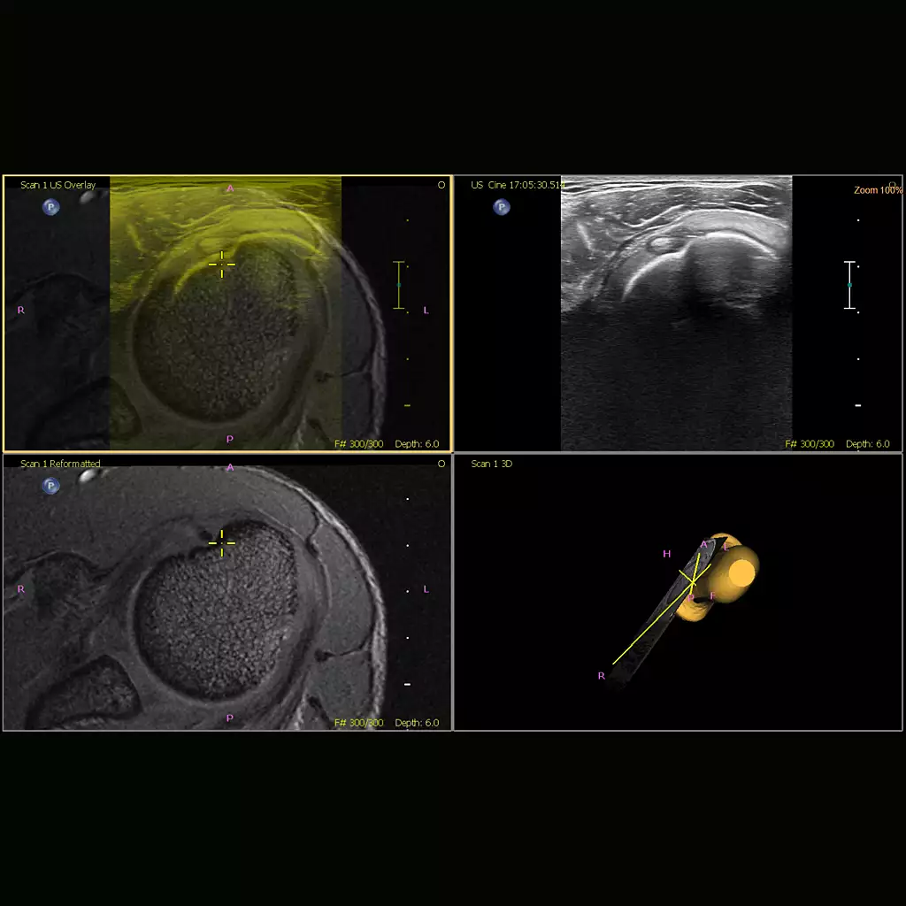
Fusion and Navigation
Make confident decisions even in challenging diagnostic cases with fully integrated fusion capabilities that feature streamlined workflows to allow clinicians to achieve fast and effective fusion of CT/MR/PET with live ultrasound. By combining imaging modalities directly on the ultrasound system, you now have access to an even more powerful diagnostic tool with advanced visualization allowing for fast clinical decisions. Expand fusion and navigation capabilities through a range of transducers across applications, including the X6-1 xMatrix , C5-1, C9-2, eL18-4, L12-5, C10-4ec, S5-1 and the new mC7-2
TrueVue advanced 3D display
Philips TrueVue advanced 3D ultrasound display delivers amazing lifelike 3D images. TrueVue, with its internal light source gives clinicians the ability to manipulate light and shadow anywhere in the 3D volume.
Auto Doppler
Automatically adjusts for optimal flow sensitivity and resolution, reducing 10 steps to 3 steps and also reducing the number of repetitive button pushes by an average of 68%.
Anatomically Intelligent ultrasound - machine intelligence for faster more reproducible analysis
At the heart of the powerful EPIQ Elite architecture is our Philips exclusive Anatomical Intelligence Ultrasound (AIUS), designed to elevate the ultrasound system from a passive to an actively adaptive device. With advanced organ modeling, image slicing, and proven quantification, exams are easy to perform, more reproducible, and deliver new levels of clinical information. Some examples of our AIUS capabilities include HeartModel, AI Breast and Auto-registration of image fusion and navigation.
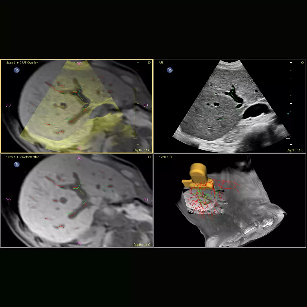
Image Fusion and Navigation-Easy to use modality fusion and interventional guidance
Make confident decisions even in challenging diagnostic cases with fully integrated fusion capabilities that feature streamlined workflows to allow clinicians to achieve fast and effective fusion of CT/MR/PET with live ultrasound. By combining imaging modalities directly on the ultrasound system, you now have access to an even more powerful diagnostic tool with advanced visualization allowing for fast clinical decisions. Expand fusion and navigation capabilities through a range of transducers across applications, including the X6-1 xMatrix, C5-1, C9-2, eL18-4, L12-5, C10-4ec, S5-1 and the new mC7-2.
Collaboration Live
Extend your team without expanding it. Collaboration Live is a communication platform that facilitates communication between a compatible ultrasound system and a remote user. With simultaneous Multi-party communication up to six users can quickly and securely talk, text, screen share and video stream directly from the ultrasound system for access to multiple clinical resources at a distance.

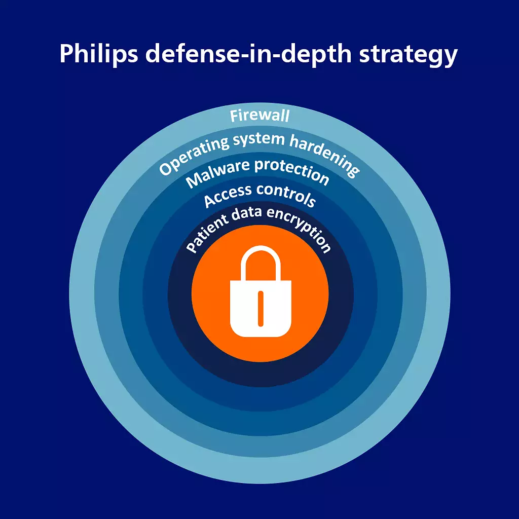
Powerful system security - protecting sensitive patient data
Hospitals and healthcare organizations are spending more to protect their systems and patient data from cyber-attacks. That is why healthcare cybersecurity spending will exceed $65 billion over the next five years. Ultrasound devices are highly mobile and can exist in a wired or wireless environment. As a result, Philips has made security a high priority for ultrasound systems. The EPIQ Elite platform is built on Window 10 OS and features a powerful defense-in-depth principle and deliver an outstanding set of data security features comprising of five core layers.
Service
The need to do more with less, rising case complexity and additional care settings put challenges with staffing, skill variability and standardization into sharp focus. This is where our services and solutions can help - get service tailored for your needs with our RightFit contracts, clinical and technical education and training to keep skills fresh, maximize your equipment investment with Technology Maximizer, and extend your team without expanding it with Collaboration Live.

Education
Our comprehensive education programs are designed to support clinical excellence, increase the use of advanced system features, instill physician confidence in the quality of exams, enhance workflow and productivity, foster professional growth and teamwork, and ultimately deliver an outstanding patient experience.

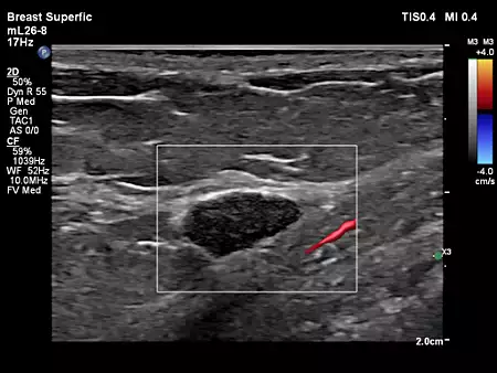
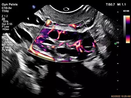
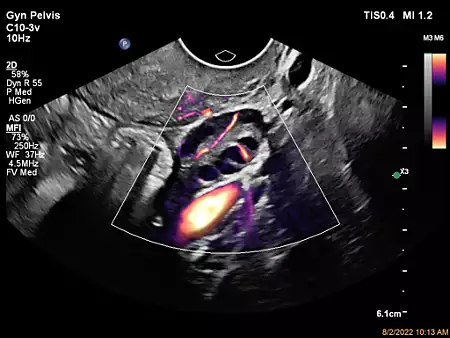
C10-3v Uterus with Flow Viewer mL26-8 Breast
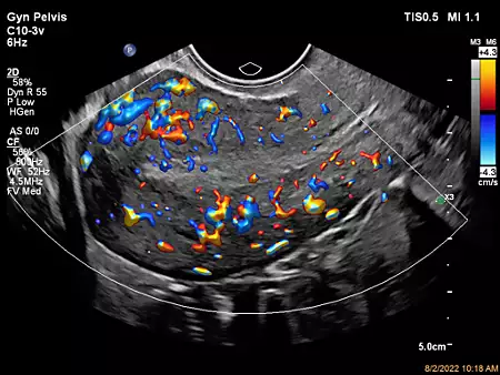
C10-3v Ovary with MicoFlow Imaging C5-1 Fetal Heart Color Flow with Flow Viewer
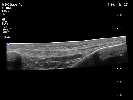
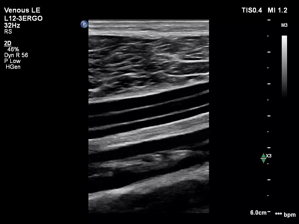
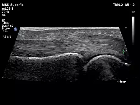
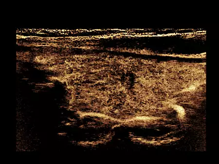
eL18-4 MSK Patella Tendon Panoramic Image L12-3ERGO Lower Limb Veins
C5-1 Renal Perfusion with MFI Flow Viewer mL26-8 MSK Wrist
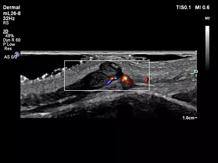
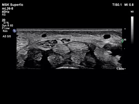
mL26-8 MSK Flexor Tendon mL26-8 Superficial Dermal Collection
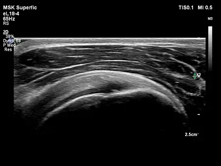
C10-3v Ovary MFI Flow Viewer eL18-4 MSK Shoulder
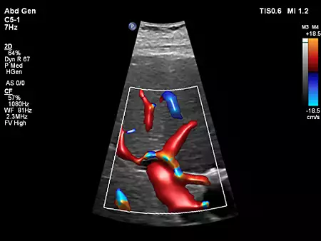
C9-2 Aortic Arch with FlowViewer eL18-4 Vertebral Perfusion with Flow Viewer

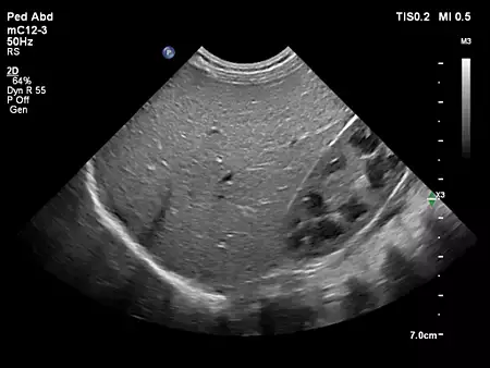
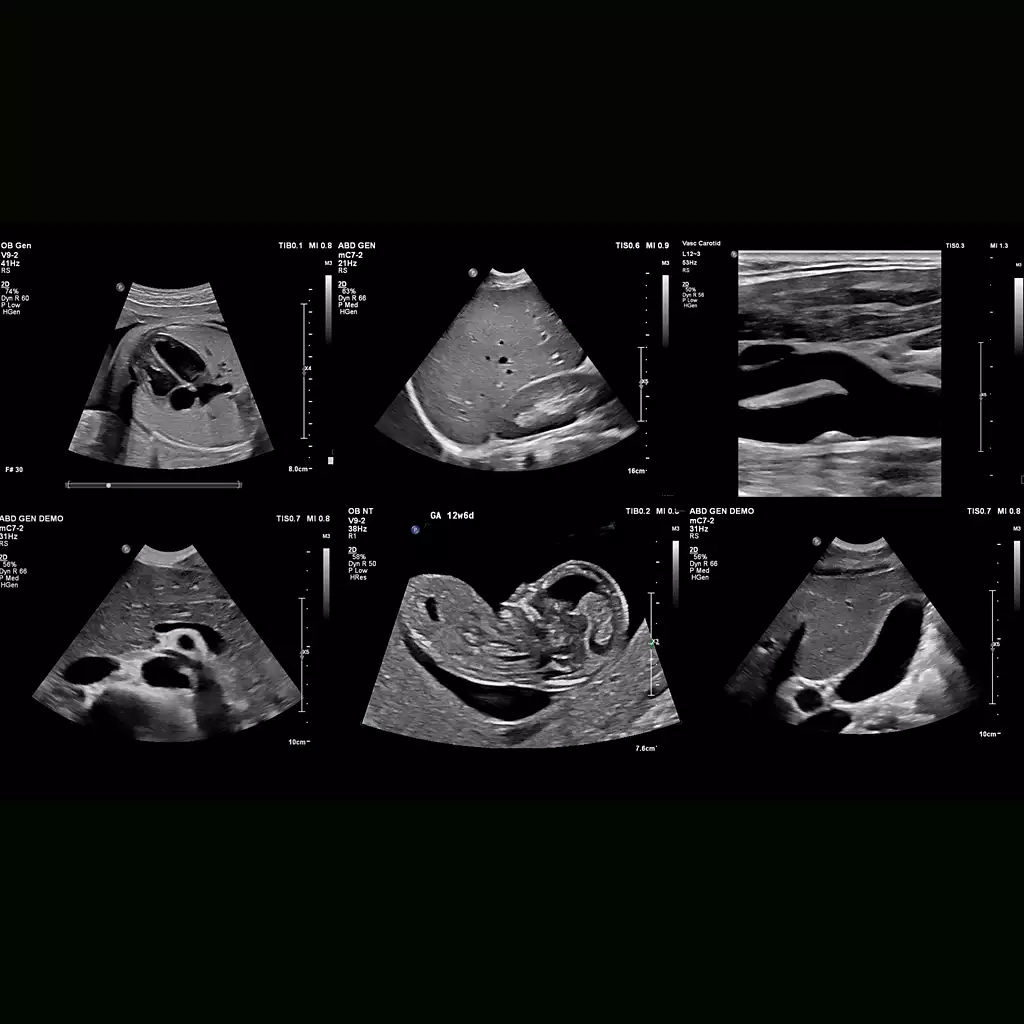
eL18-4 OB Imaging mC12-3 Pediatric Liver Kidney
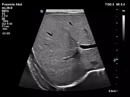
C5-1 Umbilical Cord Color Flow with Flow Viewer mL26-8 Pediatric Liver
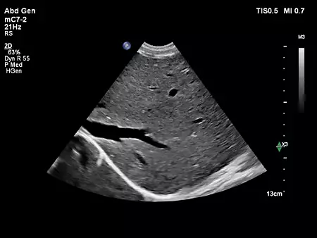
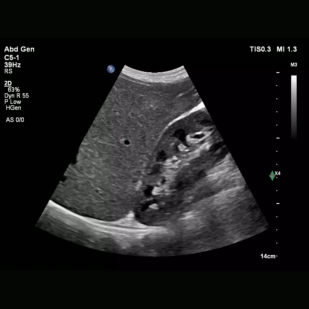
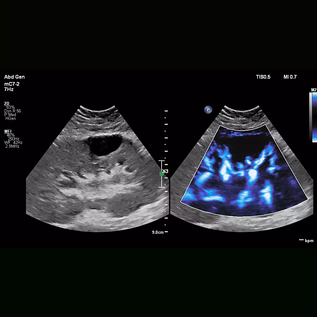
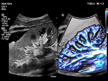
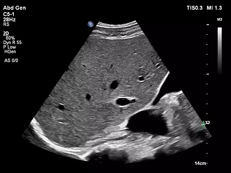
mC7-2 Abdominal Liver C5-1 Liver Perfusion with Flow Viewer
mL26-8 MSK Superficial Perfusion with MFI HD C9-2 Abdomen Liver
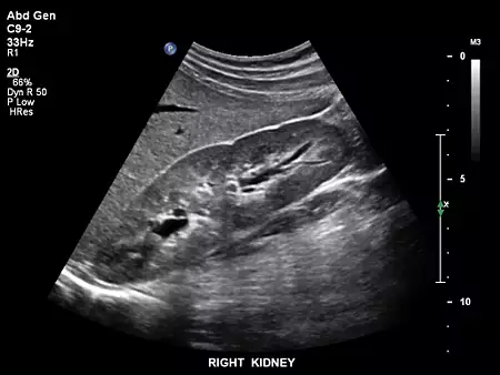
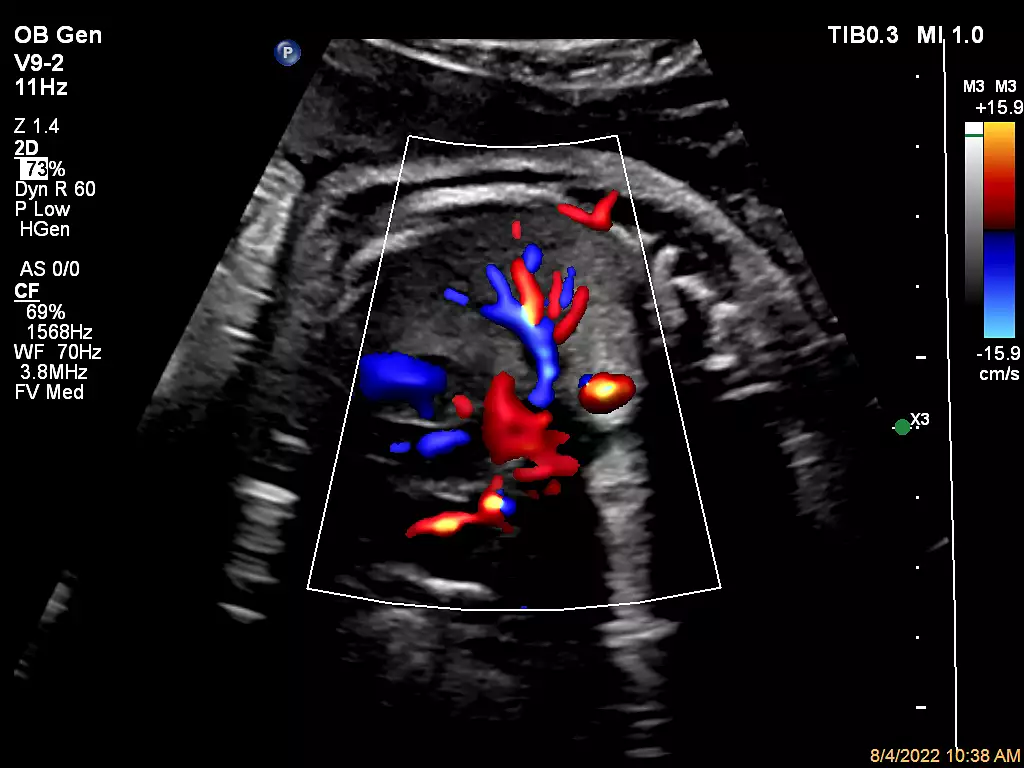
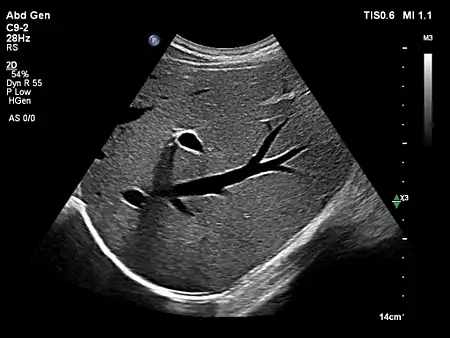
C9-2 Abdomen Renal V9-2 Fetal Lung Perfusion with Flow Viewer
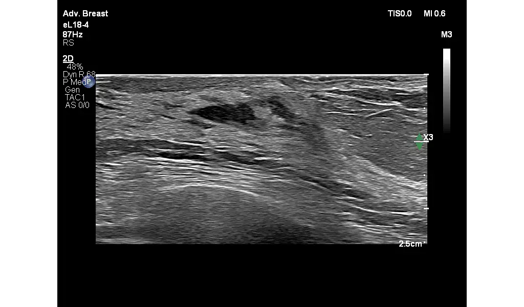
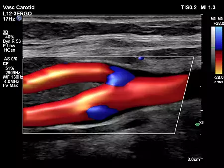
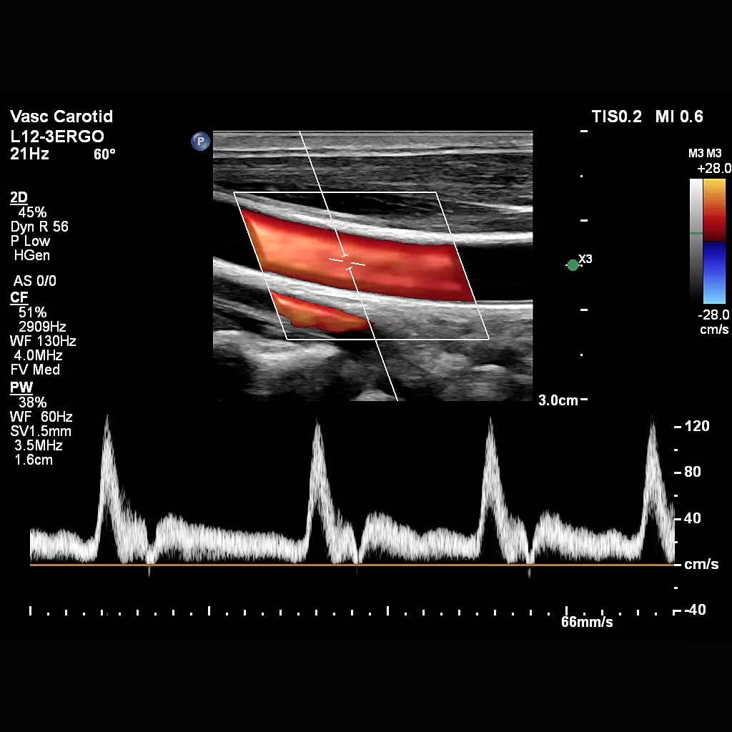
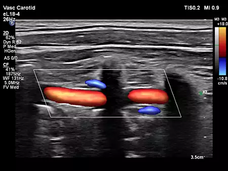
eL18-4 for Breast L12-3ERGO Vascular Carotid Flow Viewer Color
C5-1 Fetal Heart Pulmonary Vein with MFI HD (Micro Flow Imaging, High Definition) C5-1 Abdomen, with Flow Viewer
eL18-4 Testicular Imaging with MicroFlow Imaging eL18-4 Linear ElastQ Breast
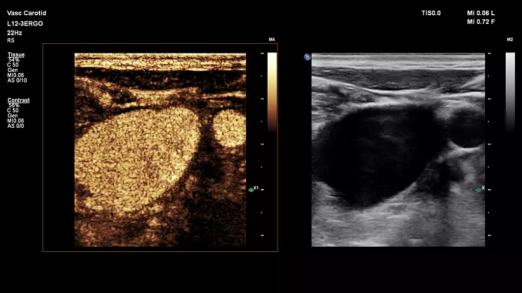
mL26-8 Vascular Fistula with Flow Viewer L12-3 paired with CEUS Auto Scan, scanning on the Vascular Carotid Preset
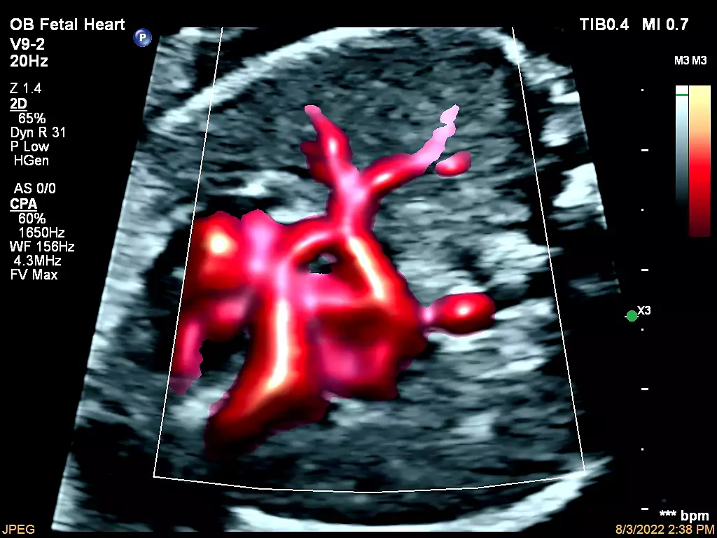
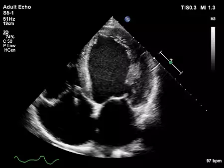
V9-2 Fetal Heart CPA with Flow Viewer S5-1 Adult Echo
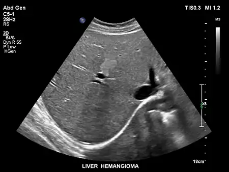
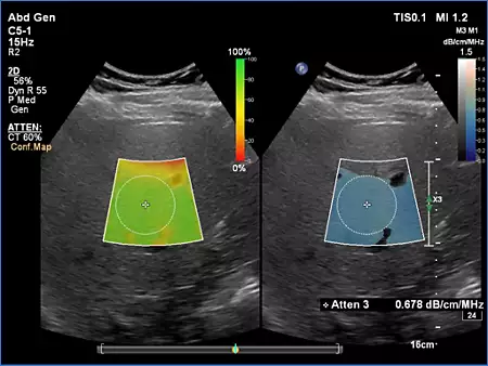
C5-1 Liver Hemangioma C5-1 Liver Fat Quantification
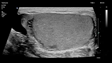
eL18-4 Testis C5-1 Abdomen
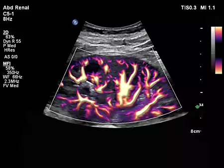
eL18-4 Thyroid CEUS C5-1 Abdomen Renal Perfusion with MFI HD
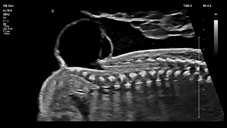
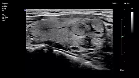
eL18-4 OB Fetal Spine eL18-4 Thyroid
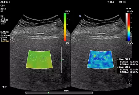
C5-1 Liver ElastQ with Confidence Map C10-3v Ovarian Perfusion with Flow Viewer
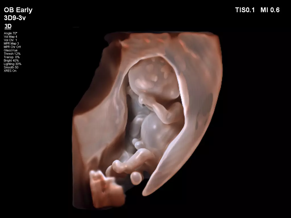
3D9-3v 3D TrueVue Fetal Imaging

 中文
中文 English