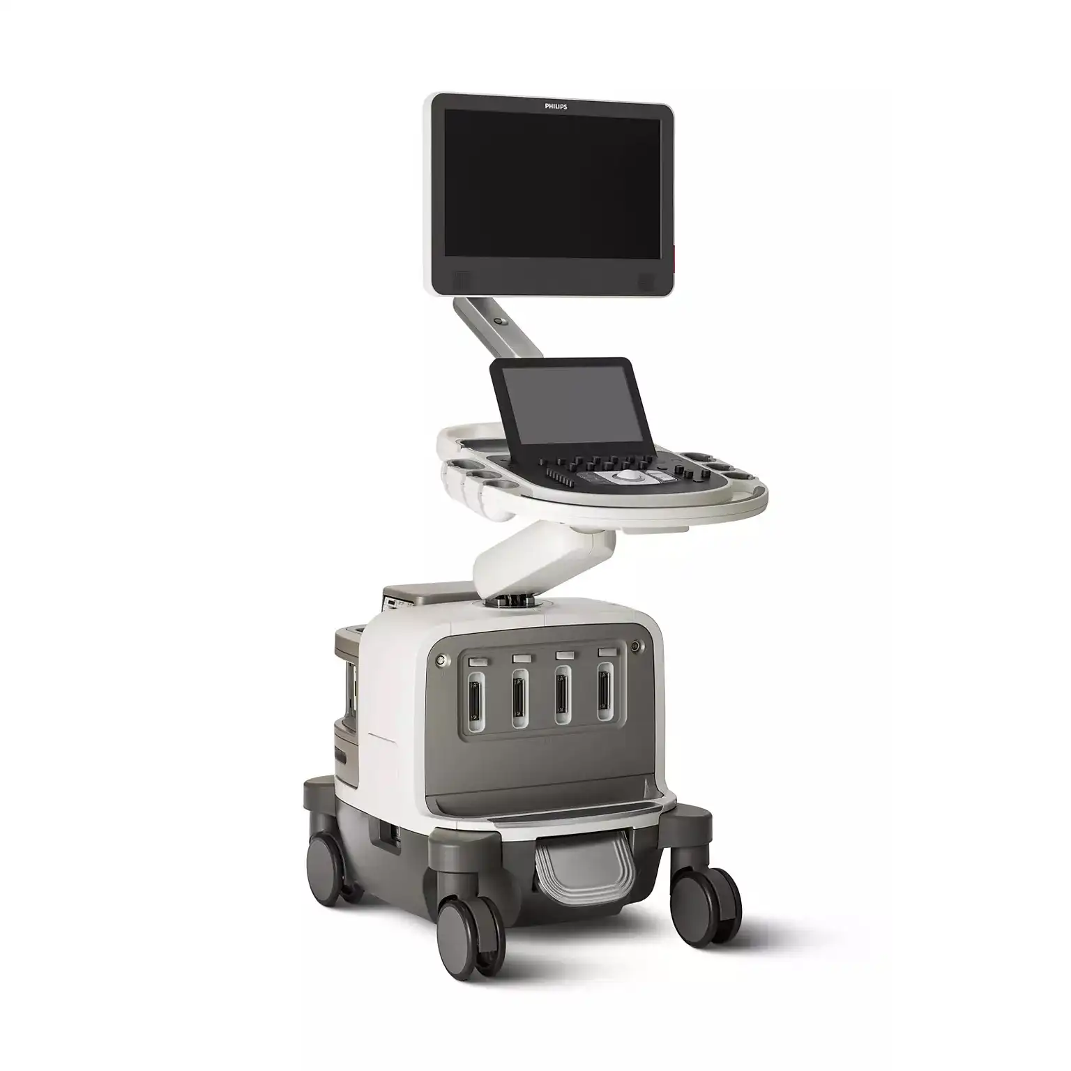
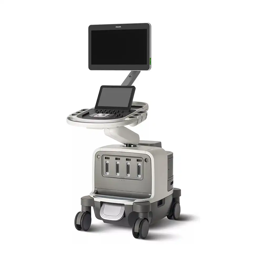
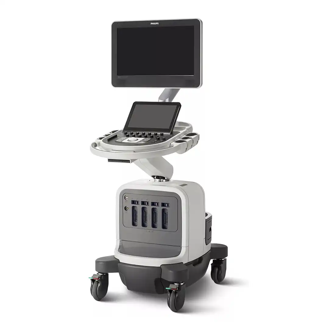
PHILIPS Affiniti 30
Ultrasound system

Choosing a new ultrasound system is all about balance. You need accurate diagnostic information quickly, a simplified yet intuitive user interface, and easy access to critical features, along with an ergonomic design and the latest technology.
Designed for balance
You go above and beyond to provide the best care for your patients. But you are expected to do so with less time, fewer resources, and higher patient volume. To balance these many demands, you need diagnostic information quickly. You need advanced functionality in an ergonomic system that is easy to use and built to last the daily rigors of high patient volume.

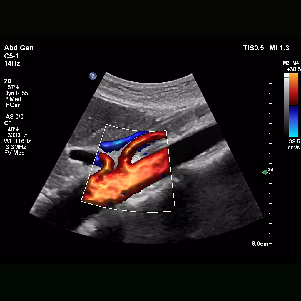
Flow Viewer
Flow Viewer is a Philips color visualization enhancement for vasculature and fetal heart architecture. Flow Viewer provides a 3D-like rendering of flow imaging data to better visualize the cardiac and vascular architecture and enhance the aesthetic appeal of all color imaging modes Available in all color imaging modes (CFM, CPA, CPAd, MFI, MFI HD).
Next Gen Auto Scan
Philips Next Gen Auto Scan improves image uniformity, adaptively adjusting image brightness at every pixel and reducing the need for user adjustment while also improving transducer plunkability. Next Gen Auto Scan reduces button pushes by up to 54% with pixel-by-pixel real-time optimization.
Workflow meets wow
With Philips Affiniti 30, workflow meets wow. The system addresses the everyday need to scan quickly and deliver results efficiently, while incorporating those innovations that make Philips ultrasound the choice of those who demand quality images and proven clinical applications.

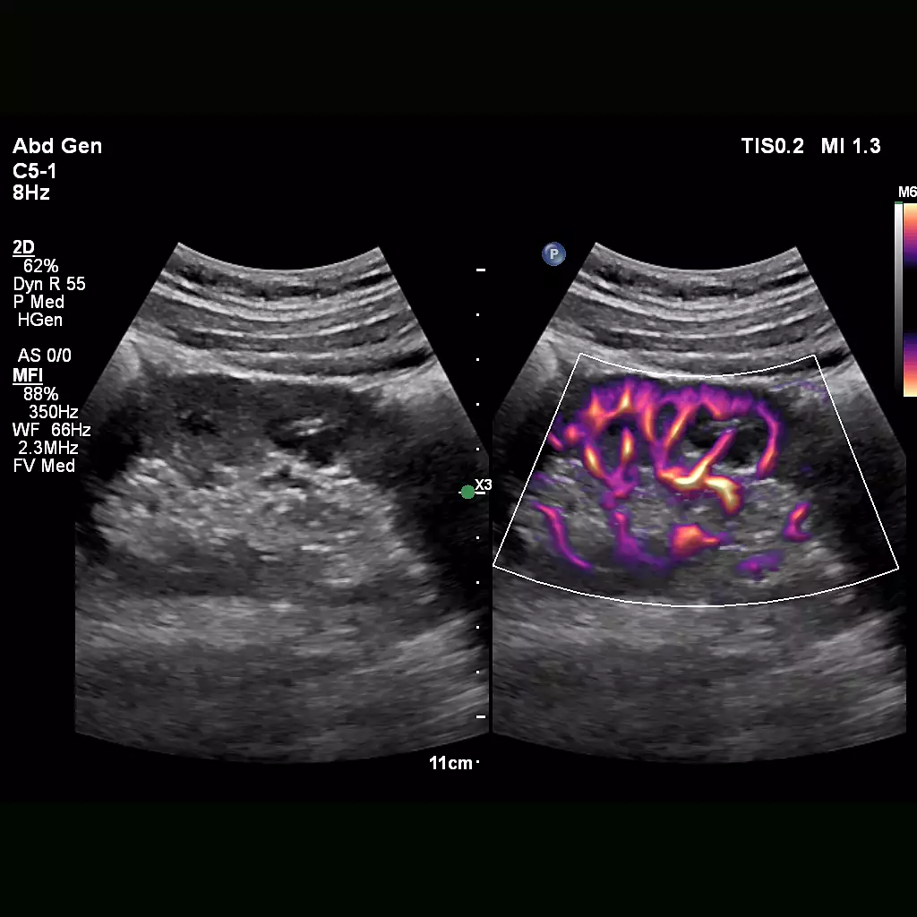
MFI
Designed to detect slow and weak blood flow anatomy in tissue. This proprietary approach overcomes many of the barriers associated with conventional methods to detect small vessel blood flow with high resolution and minimal artifacts. MicroFlow Imaging maintains high frame rate and 2D image quality while applying advanced artifact reduction techniques to reveal small vessel anatomy
Performance you can see
Affiniti 30 precision beam forming, Tissue Specific Presets (TSP), and efficiency and automation tools deliver both performance and workflow for confident throughput. Affiniti 30 provides for exceptional 3D surface rendering performance and information to diagnose fetal structures and anomalies.

Collaboration Live
Extend your team without expanding it. Collaboration Live is a communication platform that facilitates communication between a compatible ultrasound system and a remote user. With simultaneous Multi-party communication up to six users can quickly and securely talk, text, screen share and video stream directly from the ultrasound system for access to multiple clinical resources at a distance.

Comfort meets competence
Philips drew upon customer feedback when designing the Affiniti 30. We understand the challenges of daily scanning: the reality of tight spaces, high patient volume, technically difficult patients, and time constraints. We’ve designed the Affiniti 30 ultrasound machine with thoughtful details to help lighten your workload.

Service
The need to do more with less, rising case complexity and additional care settings put challenges with staffing, skill variability and standardization into sharp focus. This is where our services and solutions can help - get service tailored for your needs with our RightFit contracts, clinical and technical education and training to keep skills fresh, maximize your equipment investment with Technology Maximizer, and extend your team without expanding it with Collaboration Live.

A smart investment
The Affiniti 30 ultrasound machine boasts a low total cost of ownership, making it a smart investment. To enhance up-time, it features a modular design for enhanced reliability and rapid repair. In addition, Philips remote services* monitoring, which corrects issues using a standard Internet connection, reducing the need for service calls 3) Access to our award-winning service organization.

Education
Our comprehensive education programs are designed to support clinical excellence, increase the use of advanced system features, instill physician confidence in the quality of exams, enhance workflow and productivity, foster professional growth and teamwork, and ultimately deliver an outstanding patient experience.

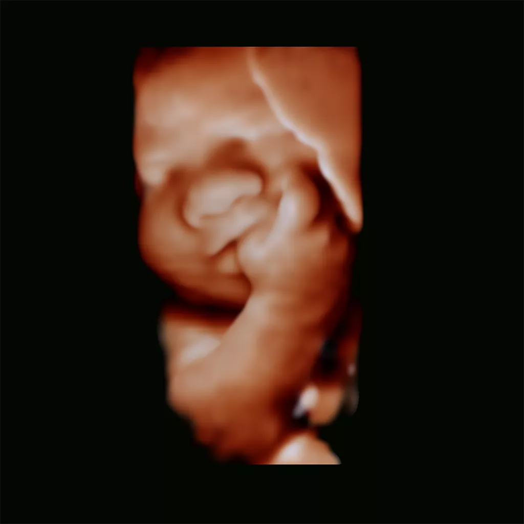
Advanced 3D visualization feature
Philips suite of advanced visualization features now available on Affiniti 30. These allow you to easily acquire lifelike 3D images. Philips TrueVue and GlassVue, with internal light source, deliver photorealistic fetal images. aReveal automatic 3D segmentation allows you to reveal the fetal face with one touch. Philips TrueVue advanced 3D ultrasound display delivers amazing lifelike 3D images. TrueVue, with its internal light source gives clinicians the ability to manipulate light and shadow anywhere in the 3D volume.
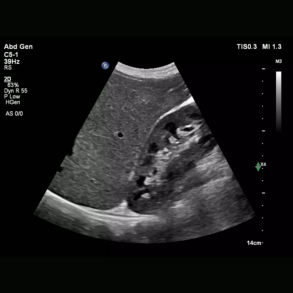
Clinical image gallery
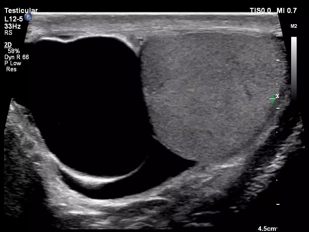
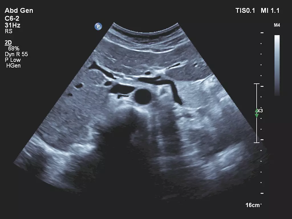
L12-5 Testicular Imaging C6-2 Abdominal Imaging
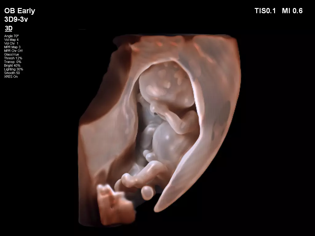
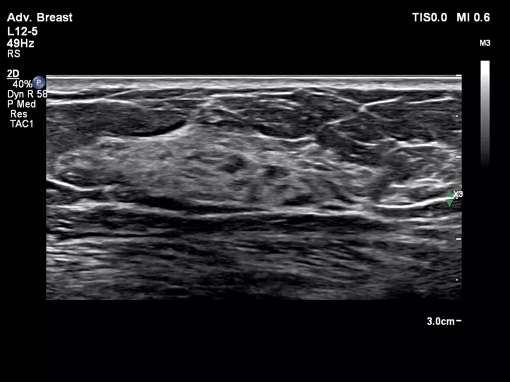
3D9-3v Fetal Imaging with TrueVue L12-5 Advanced Breast
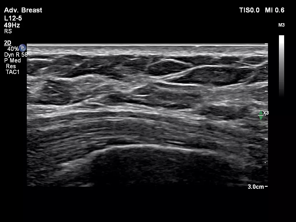
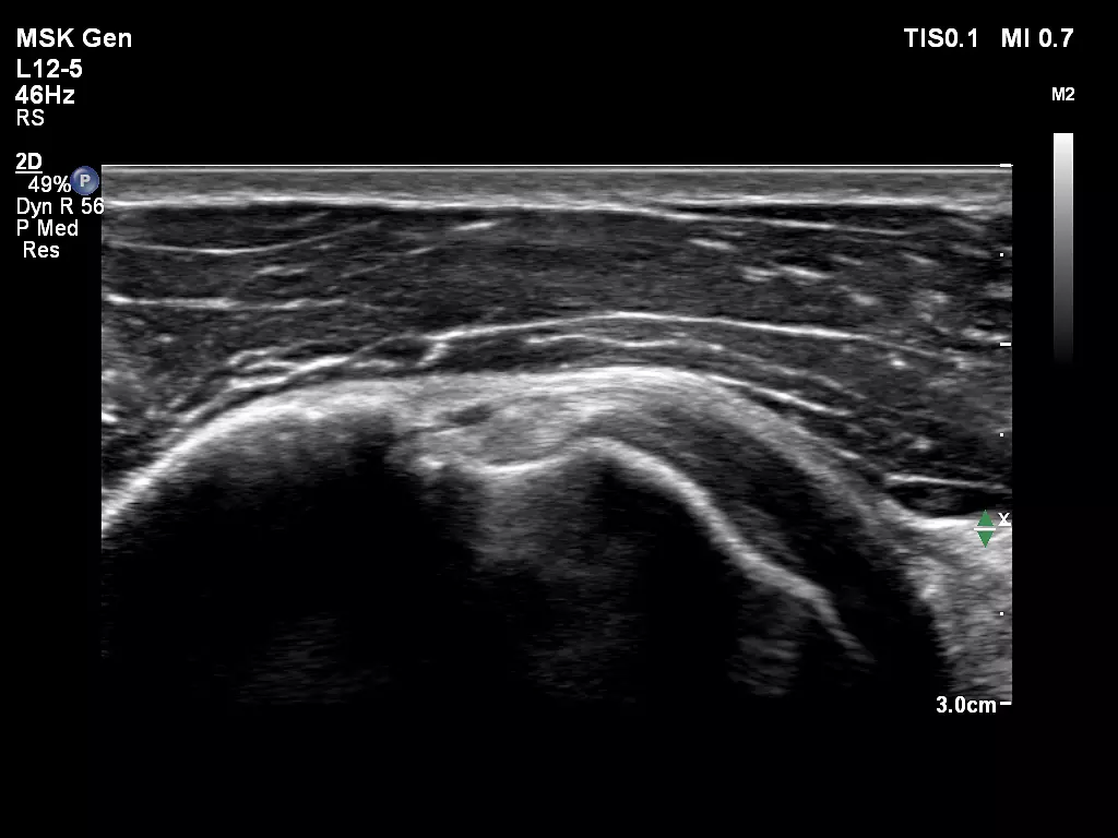
L12-5 Advanced Breast L12-5 MSK Biceps Tendon
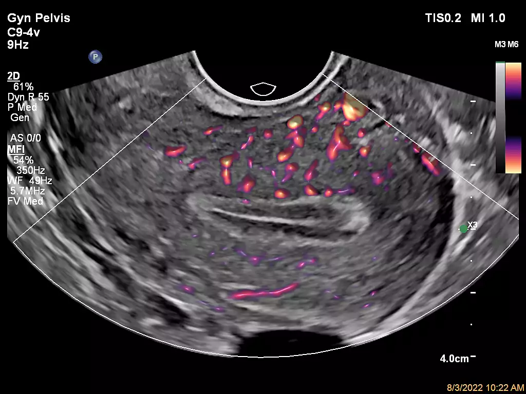
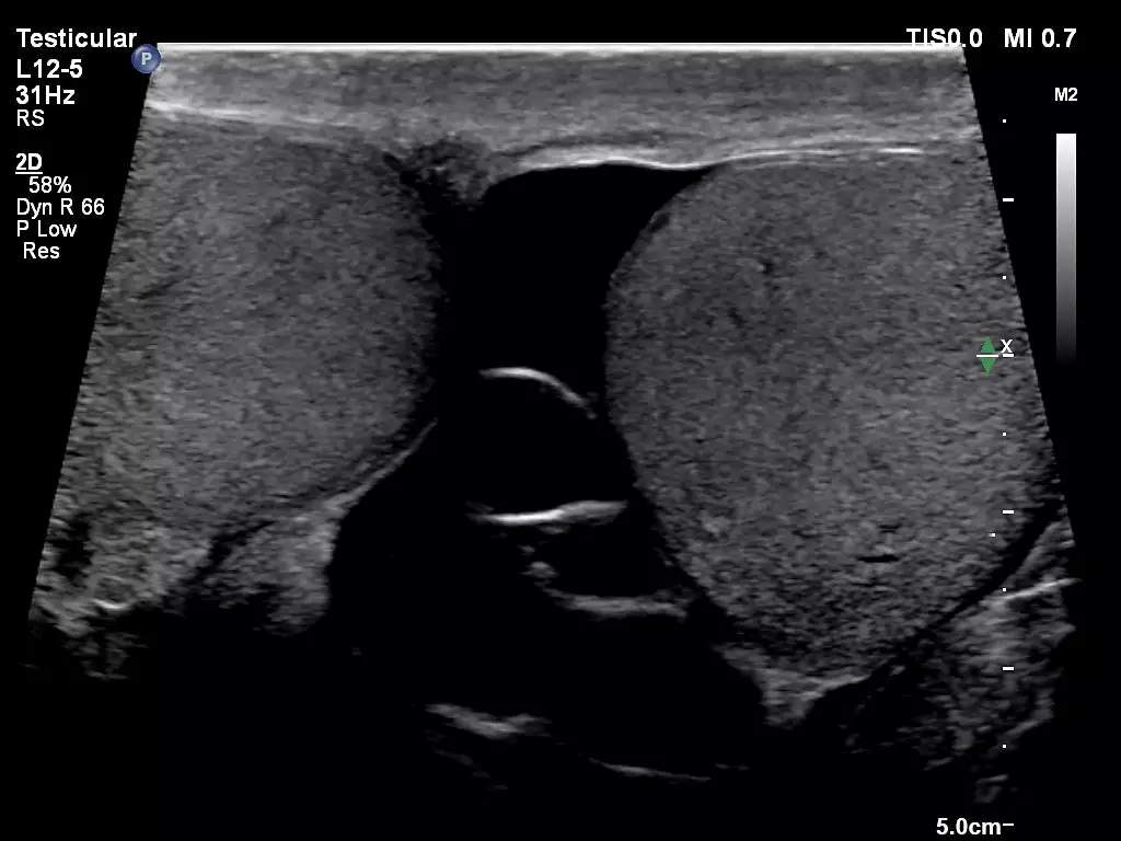
C9-4v Gynecology with MFI L12-5 Testicular Imaging
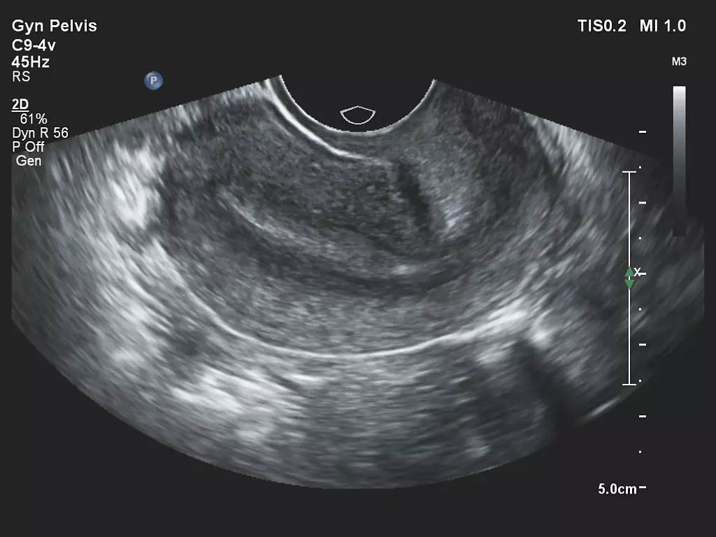
C9-4v Gynecology Imaging


 Microsystem
 Endoscopysystem
 Energysystem
 EndoscopyConsumables
 +86-21-54286005
 Room 602, Building 1, No. 111 Luxiang Road (Greenland Park Plaza), Baoshan District, Shanghai, China
  English
 中文
中文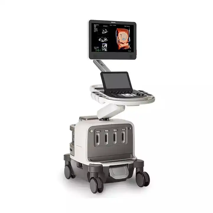
 Affiniti 30_Brochure_EN


 中文
中文 English