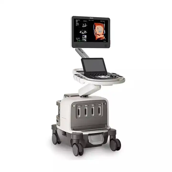
PHILIPS Affiniti 50
Ultrasound system

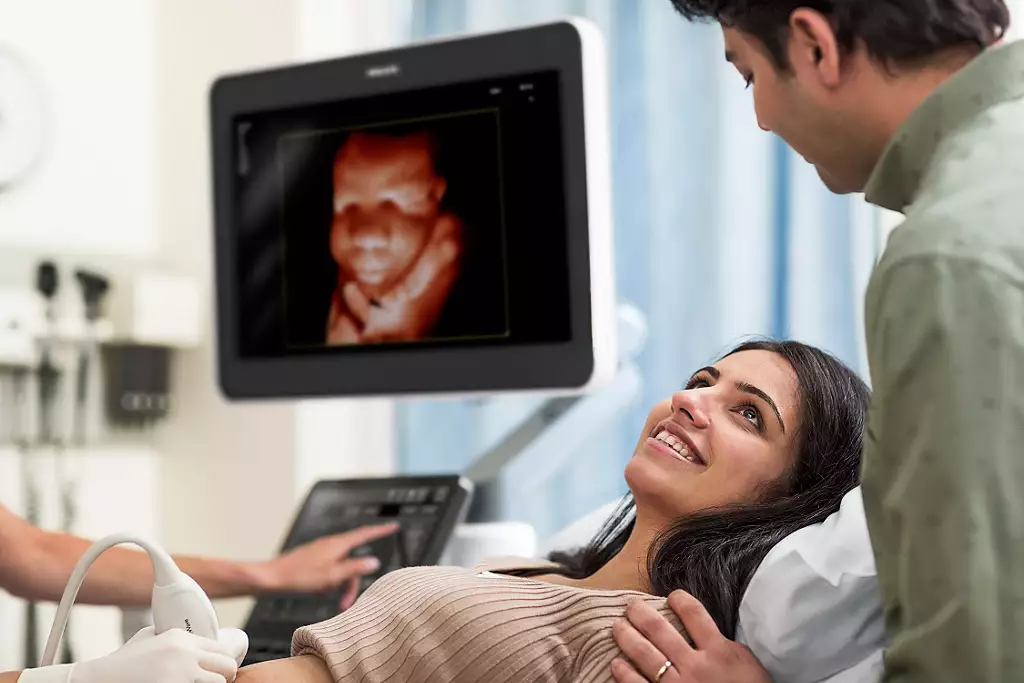
Affiniti 50 Elevate provides stunning imaging and exceptional value, delivering versatile clinical capabilities and reproducibility with ease. With streamlined workflow and reliable performance, it helps you deliver the best possible care every day
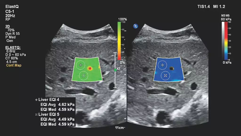
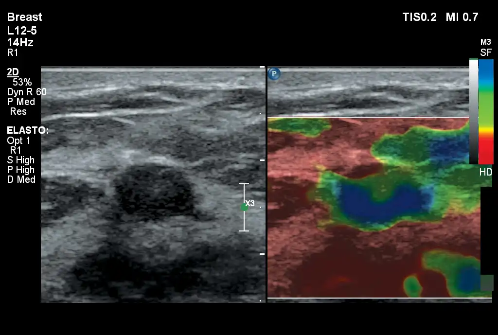
Auto ElastQ
Perform automated liver elastography with Auto ElastQ, and experience our next generation of liver health assessment. Auto ElastQ is designed to simplify user workflow with real-time, quantitative shear wave measurements.
Contrast-enhanced ultrasound (CEUS)
CEUS can transform the role of ultrasound in the liver, allowing the study of the enhancement patterns of suspicious liver lesions in real time, as well as provide an alternative non-ionizing approach to the assessment of vesicoureteral reflux in pediatric patients.
_EN.webp)
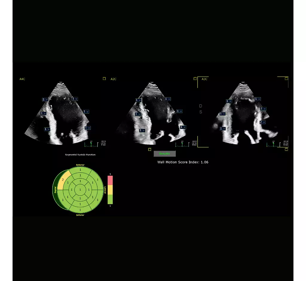
Auto Segmental Wall Motion Scoring
Provides automated evaluation of wall motion in a standard 17-segment bullseye display to aid objective LV wall assessment. With Auto SWMS, you can achieve greater reproducibility and efficiency in your workflows.
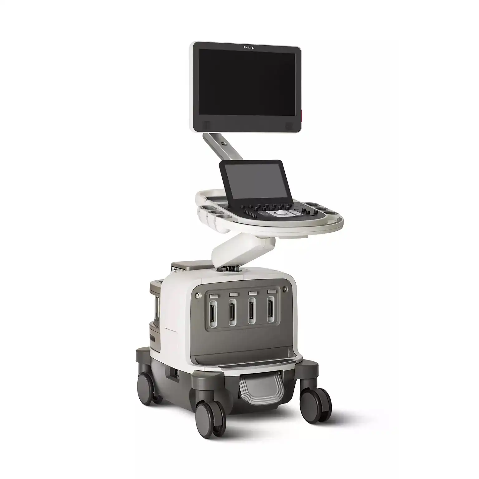
It understands your everyday
Philips Affiniti delivers the right balance of advanced ergonomic design and precision engineering to help you work more comfortably and intuitively. Its exceptional image quality gives you the results you need to provide the best patient care possible. See what Philips Affiniti can do for your practice.
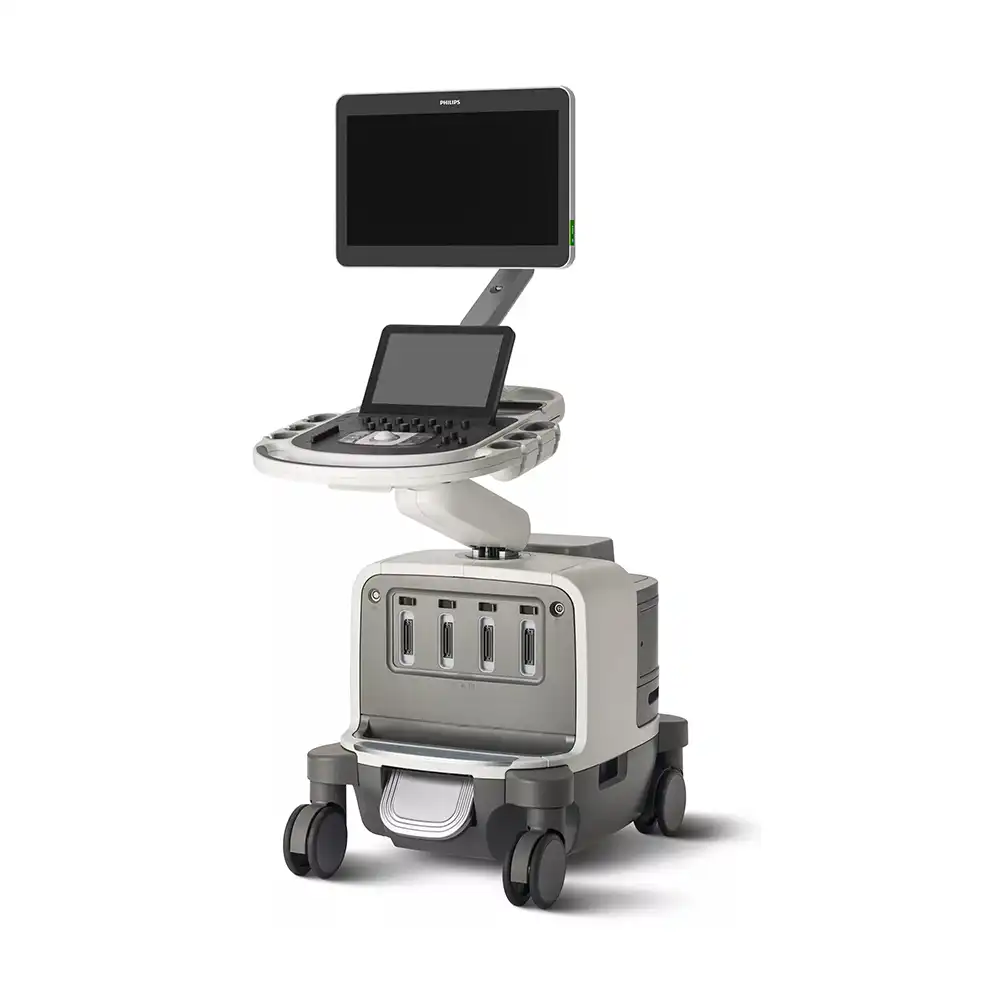
Designed for balance
You go above and beyond to provide the best care for your patients. But you are expected to do so with less time, fewer resources, and higher patient volume. To balance these many demands, you need diagnostic information quickly. You need advanced functionality in an ergonomic system that is easy to use and built to last the daily rigors of high patient volume.
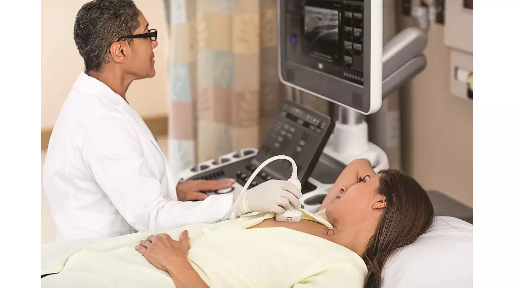
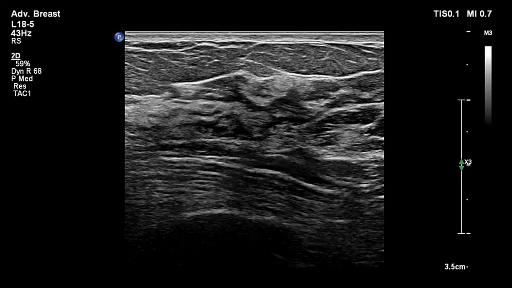
Anatomical Intelligence for Breast
Philips AI Breast is an integrated solution for whole breast ultrasound. AI Breast offers screening, diagnostic, and workflow benefits utilizing Philips unique Anatomical Intelligence. Designed with both the user and patient in mind, AI Breast allows the ultrasound scan room to be utilized for a full range of examinations without additional obtrusive hardware.
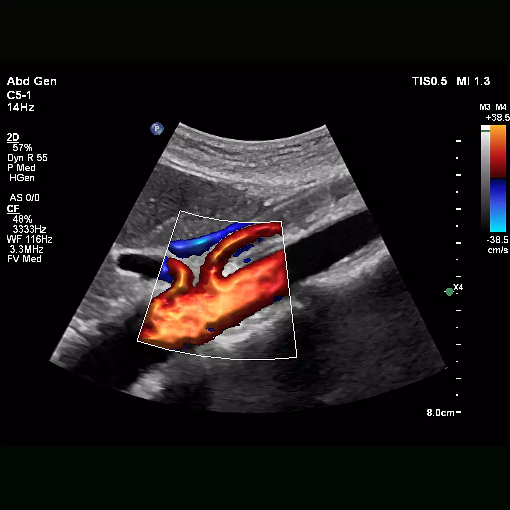
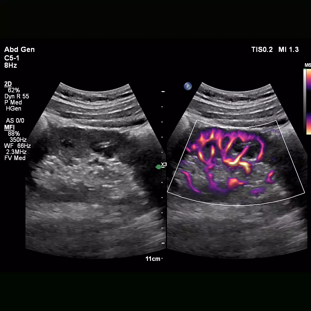
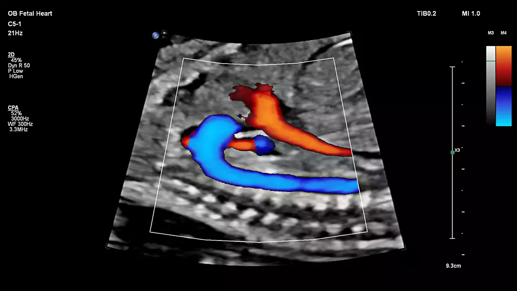
Flow Viewer
Flow Viewer is a Philips color visualization enhancement for vasculature and fetal heart architecture. Flow Viewer provides a 3D-like rendering of flow imaging data to better visualize the cardiac and vascular architecture and enhance the aesthetic appeal of all color imaging modes Available in all color imaging modes (CFM, CPA, CPAd, MFI, MFI HD).
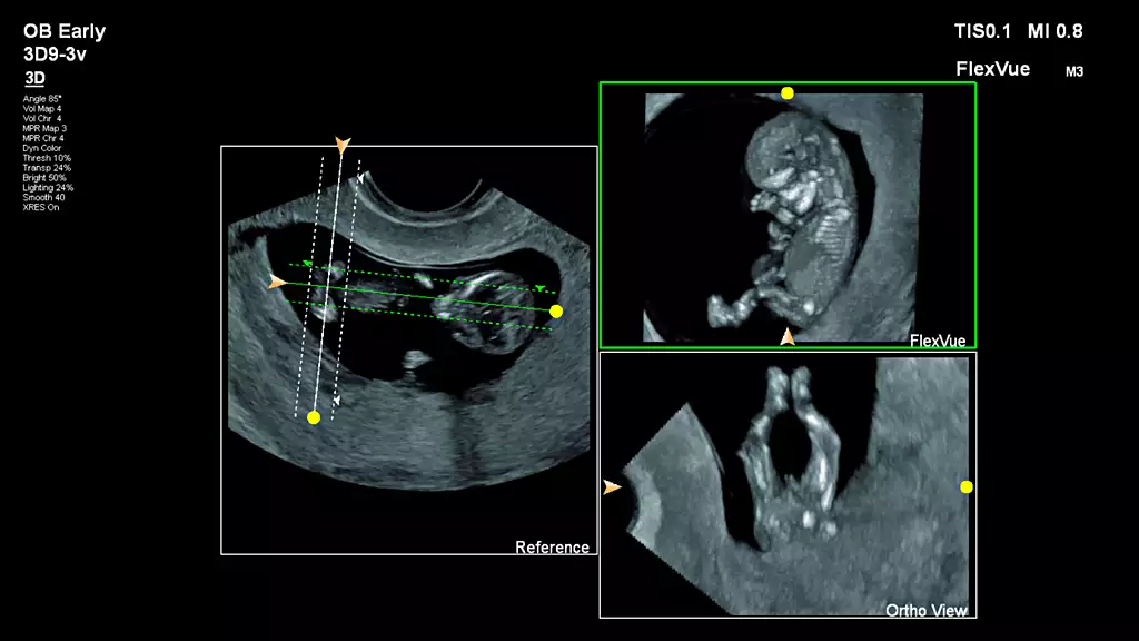
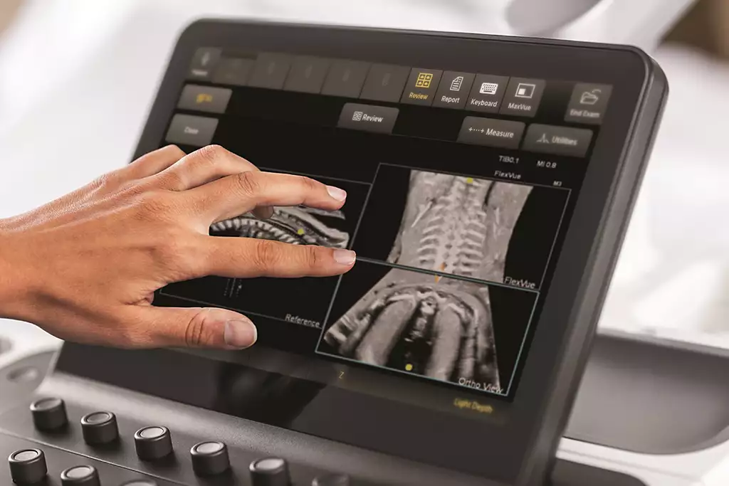
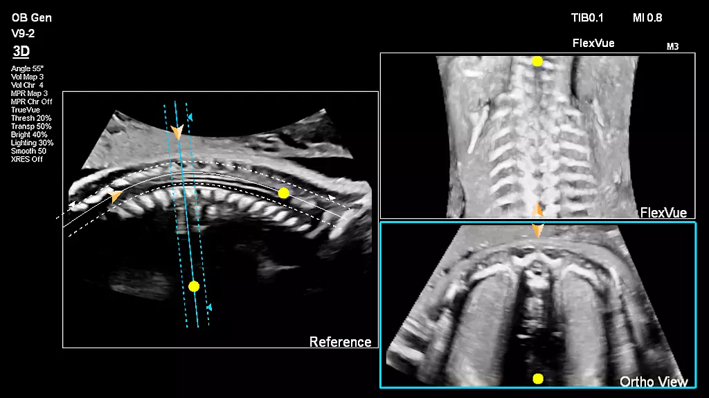
FlexVue with Orthogonal View
Easy-to-use tools designed to extract challenging anatomical planes from 3D data sets. This advanced feature offers exceptional flexibility in plane acquisition, complemented by a comprehensive measurement package for precise quantification.
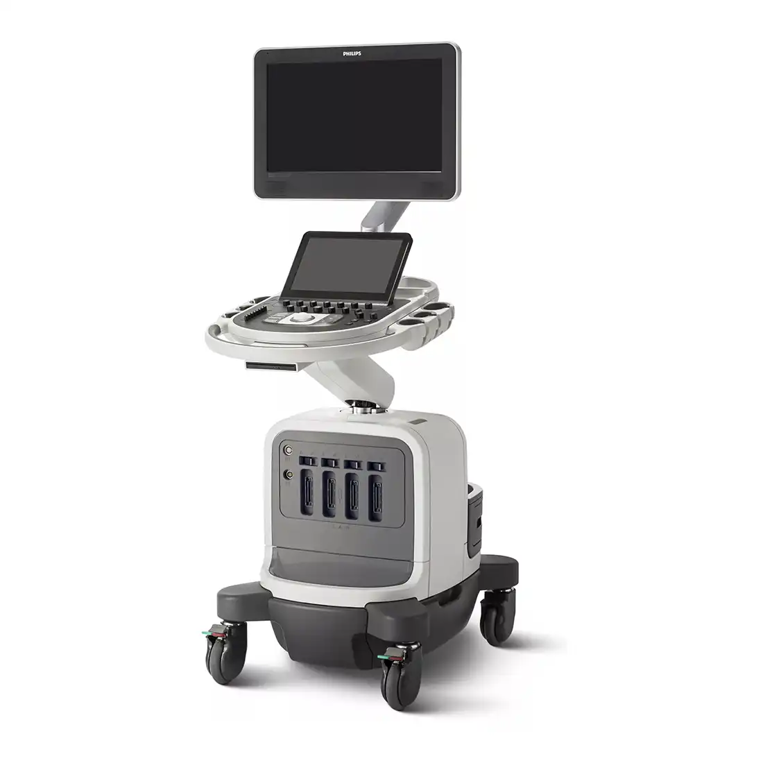
Workflow meets wow
With Philips Affiniti 50, workflow meets wow. The system addresses the everyday need to scan quickly and deliver results efficiently, while incorporating those innovations that make Philips ultrasound the choice of those who demand quality images and proven clinical applications.
Next Gen Auto Scan
Philips Next Gen Auto Scan improves image uniformity, adaptively adjusting image brightness at every pixel and reducing the need for user adjustment while also improving transducer plunkability. Next Gen Auto Scan reduces button pushes by up to 54% with pixel-by-pixel real-time optimization.
Performance you can see
Affiniti 50’s precision beamforming, Tissue Specific Presets (TSP), and efficiency and automation tools deliver both performance and workflow for confident throughput. The system’s outstanding image quality combines with advanced clinical functionality, including elastography and Anatomical Intelligence Ultrasound (AIUS).
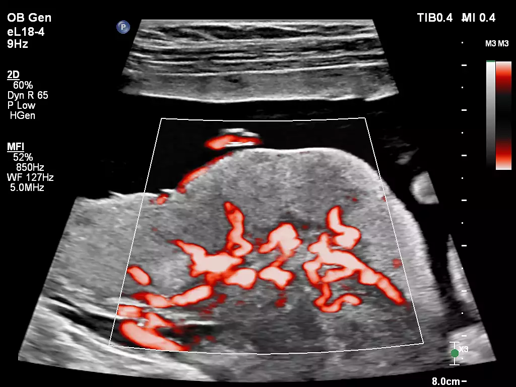
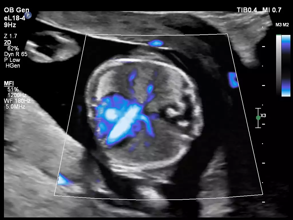
MFI
Designed to detect slow and weak blood flow anatomy in tissue. This proprietary approach overcomes many of the barriers associated with conventional methods to detect small vessel blood flow with high resolution and minimal artifacts. MicroFlow Imaging maintains high frame rate and 2D image quality while applying advanced artifact reduction techniques to reveal small vessel anatomy
Collaboration Live
Extend your team without expanding it. Collaboration Live is a communication platform that facilitates communication between a compatible ultrasound system and a remote user. With simultaneous Multi-party communication up to six users can quickly and securely talk, text, screen share and video stream directly from the ultrasound system for access to multiple clinical resources at a distance.

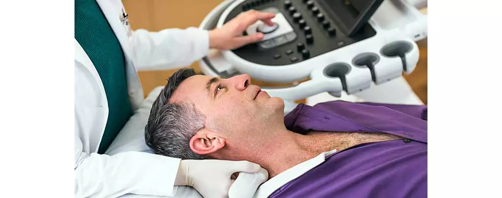
Comfort meets competence
Philips leverages the experiences of its customers to design Affiniti 50 to address the challenges of daily scanning. We understand the reality of tight spaces, high patient volume, technically difficult patients and time constraints, and we’ve designed the system with thoughtful details to help lighten your workload.

A smart investment
The Affiniti 50 boasts a low total cost of ownership, making it a smart investment. To enhance uptime, it features: 1) A modular design for enhanced reliability and rapid repair 2) Philips remote services* monitoring, which corrects issues using a standard Internet connection, reducing the need for service calls 3) Access to our award-winning service organization.

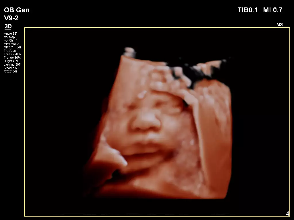
TrueVue advanced 3D display
Philips TrueVue advanced 3D ultrasound display delivers amazing lifelike 3D images. TrueVue, with its internal light source gives clinicians the ability to manipulate light and shadow anywhere in the 3D volume.
MaxVue high definition display
At the touch of a button, MaxVue high-definition display brings extraordinary visualization of anatomy with 1,179,648 additional image pixels compared to a standard 4:3 display format mode. MaxVue enhances ultrasound viewing and provides 38% more viewing area to optimize the display of dual, side/side, biplane, and scrolling imaging modes.
Service
The need to do more with less, rising case complexity and additional care settings put challenges with staffing, skill variability and standardization into sharp focus. This is where our services and solutions can help - get service tailored for your needs with our RightFit contracts, clinical and technical education and training to keep skills fresh, maximize your equipment investment with Technology Maximizer, and extend your team without expanding it with Collaboration Live.

Education
Our comprehensive education programs are designed to support clinical excellence, increase the use of advanced system features, instill physician confidence in the quality of exams, enhance workflow and productivity, foster professional growth and teamwork, and ultimately deliver an outstanding patient experience.

Clinical image gallery
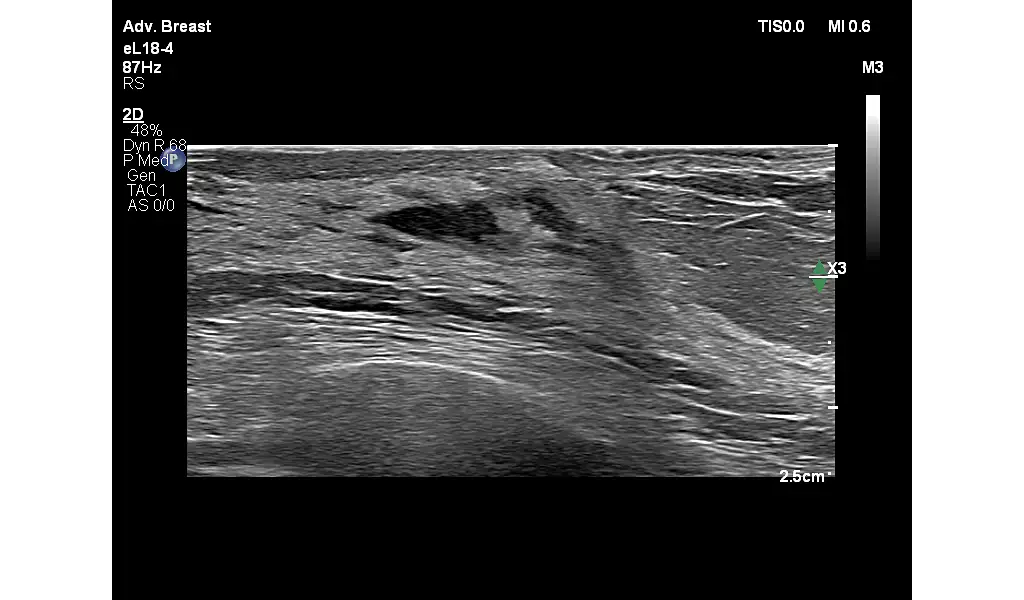
eL18-4 for Breast
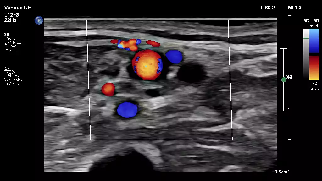
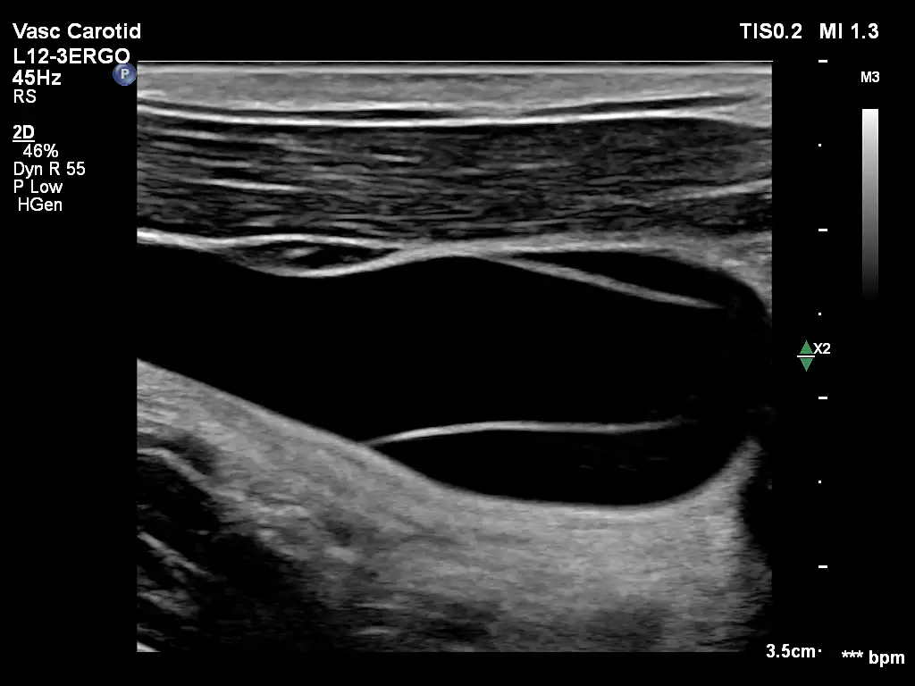
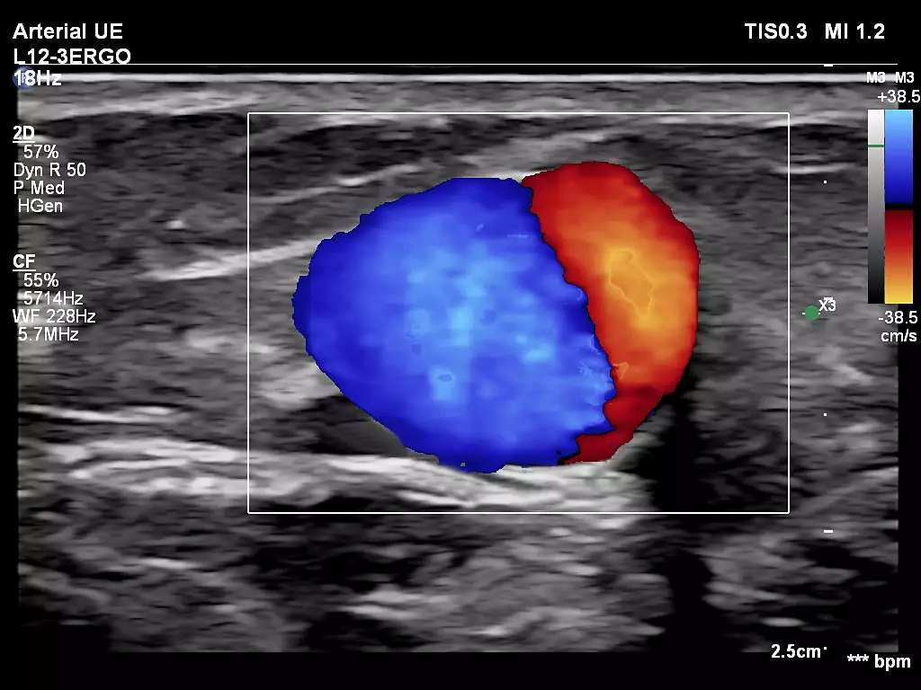
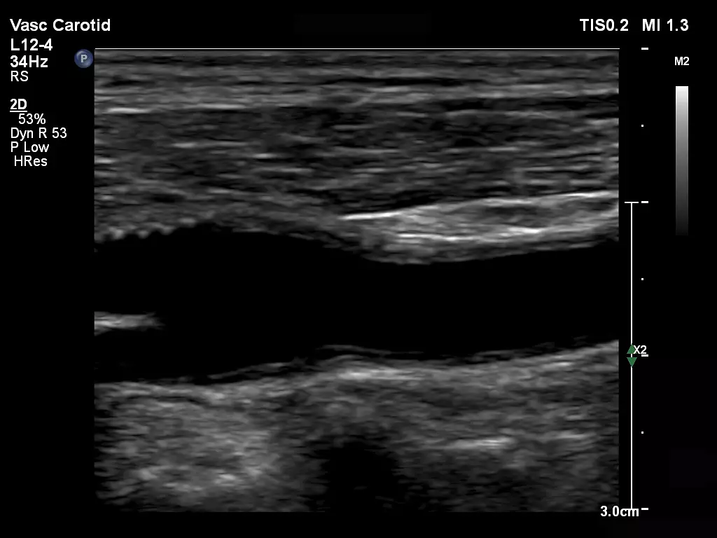
L12-3ERGO Vascular Carotid Bulb
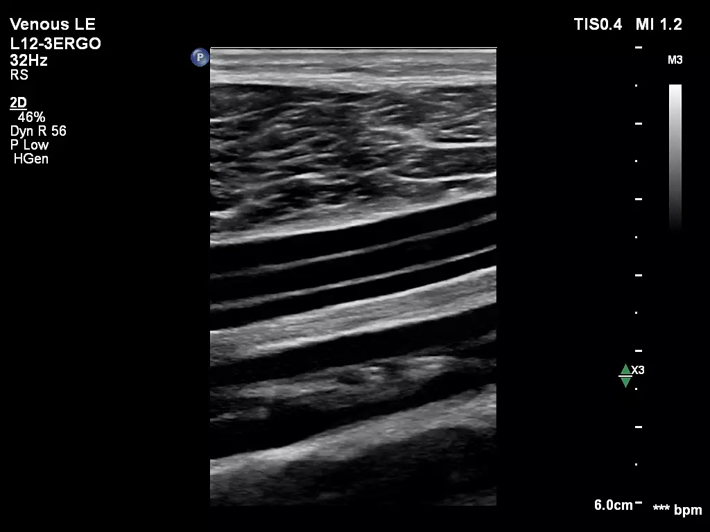
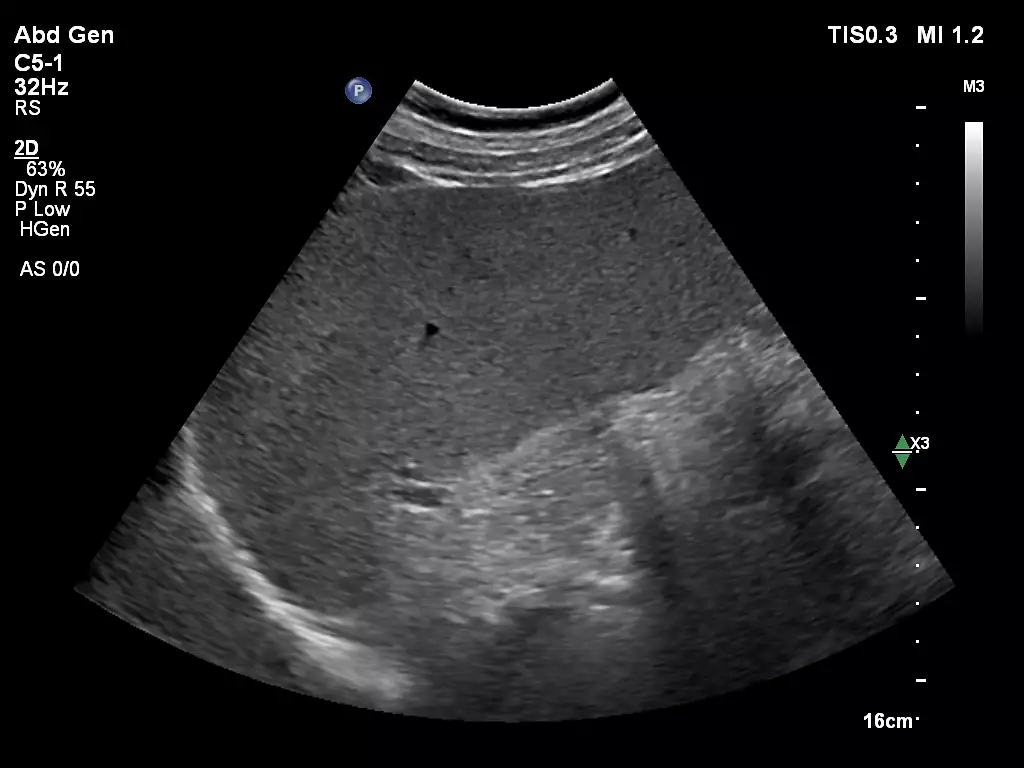
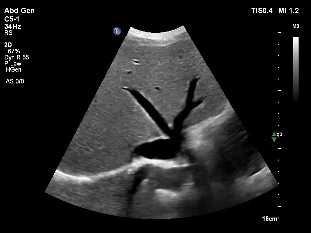
L12-3ERGO Calf Veins C5-1 Abdomen Spleen Imaging
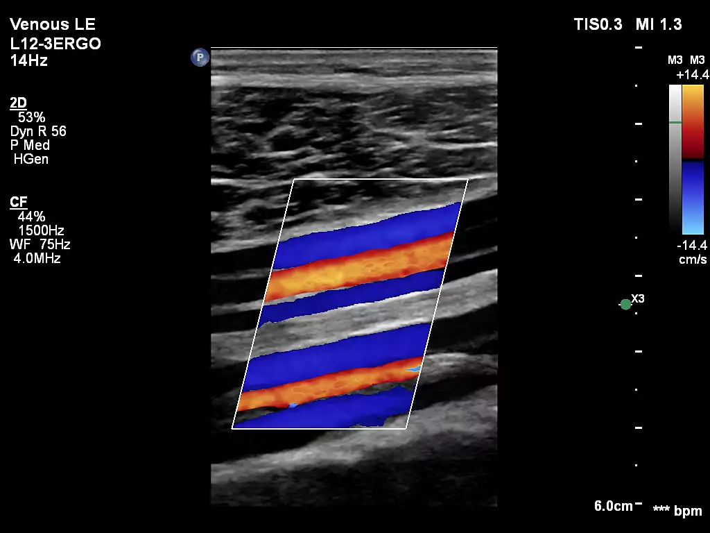
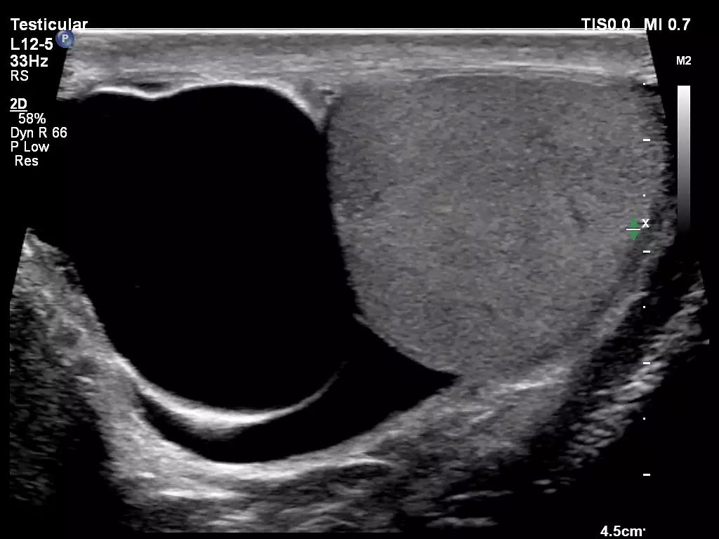
mL26-8 paired with Flow Viewer, scanning on a calf vein L12-5 Testicular Imaging
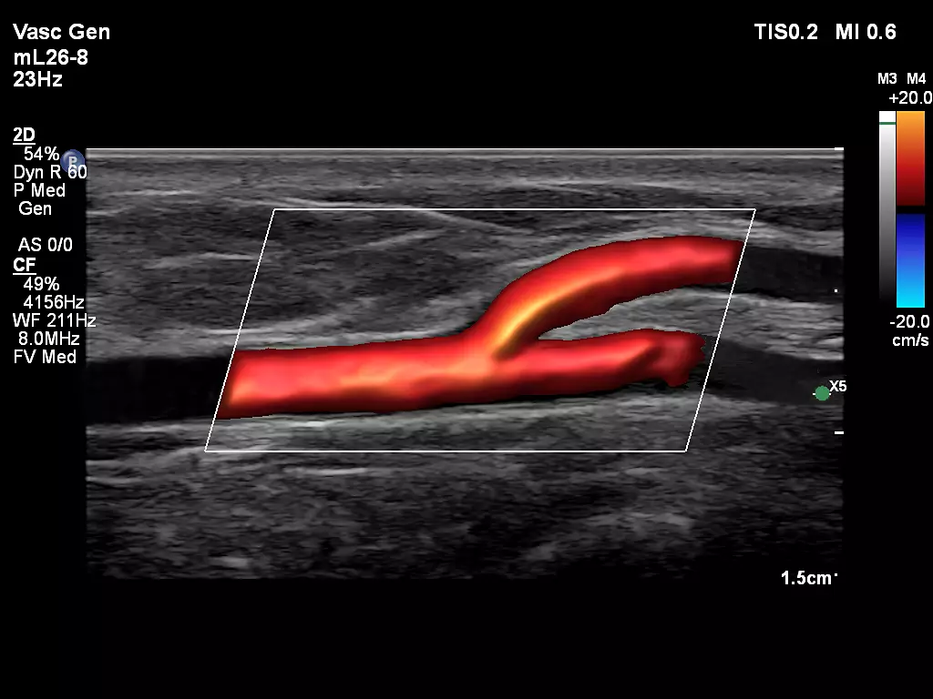
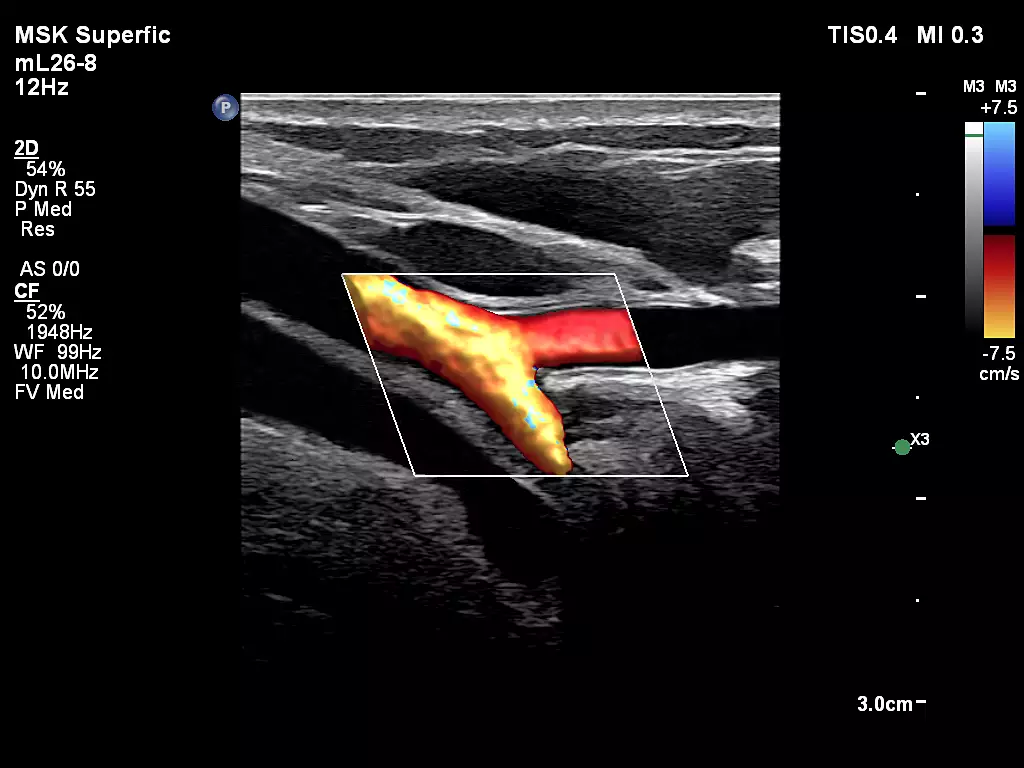
mL26-8 Superficial Artery with FlowViewer C5-1 Abdominal Imaging
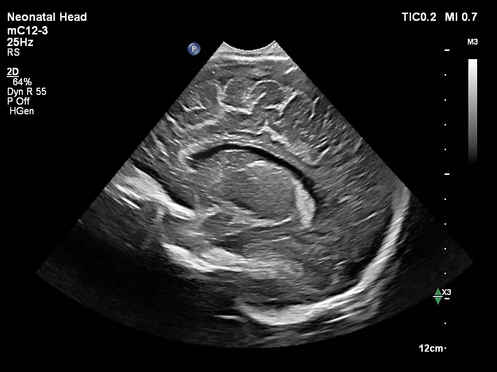
L12-3ERGO Internal Jugular Vein mC12-3 Neonatal Head
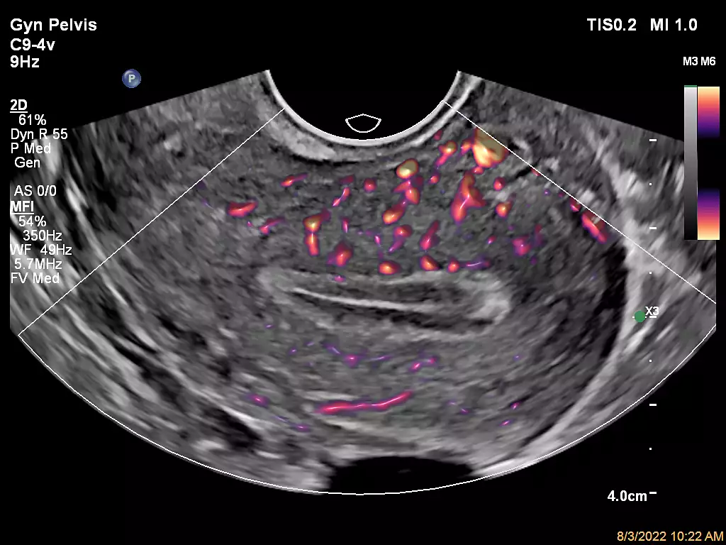
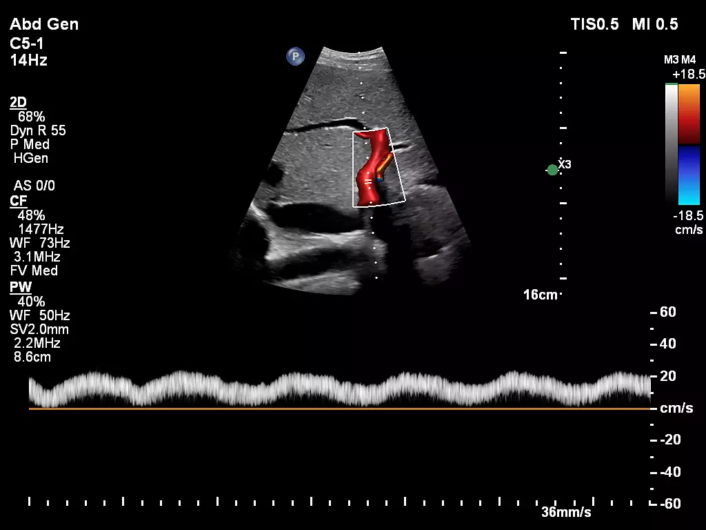
C9-4v Gynecology with MFI C5-1 Liver Doppler with FlowViewer
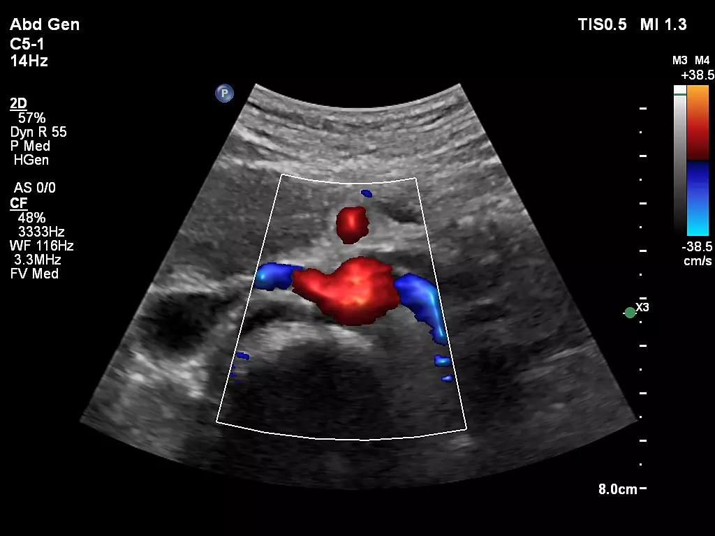
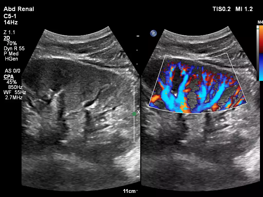
L12-3ERGO Fistula Imaging C5-1 Renal Artery Origin with FlowViewer
_EN.webp)
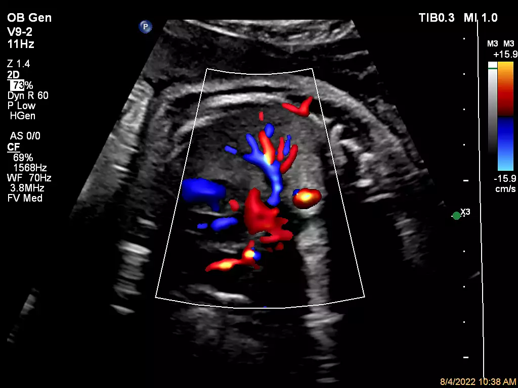
V9-2 Fetal Lung Perfusion with Flow Viewer C5-1 Hepatic Renal Index (HRI)
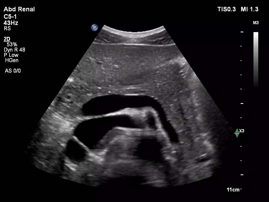
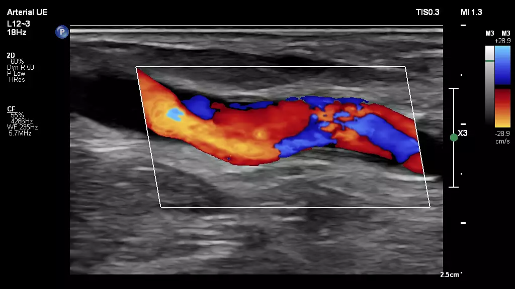
C5-1 Renal Kidney Imaging mL26-8 Upper Extremity Arterial
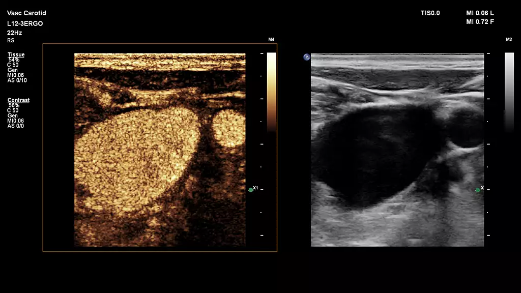
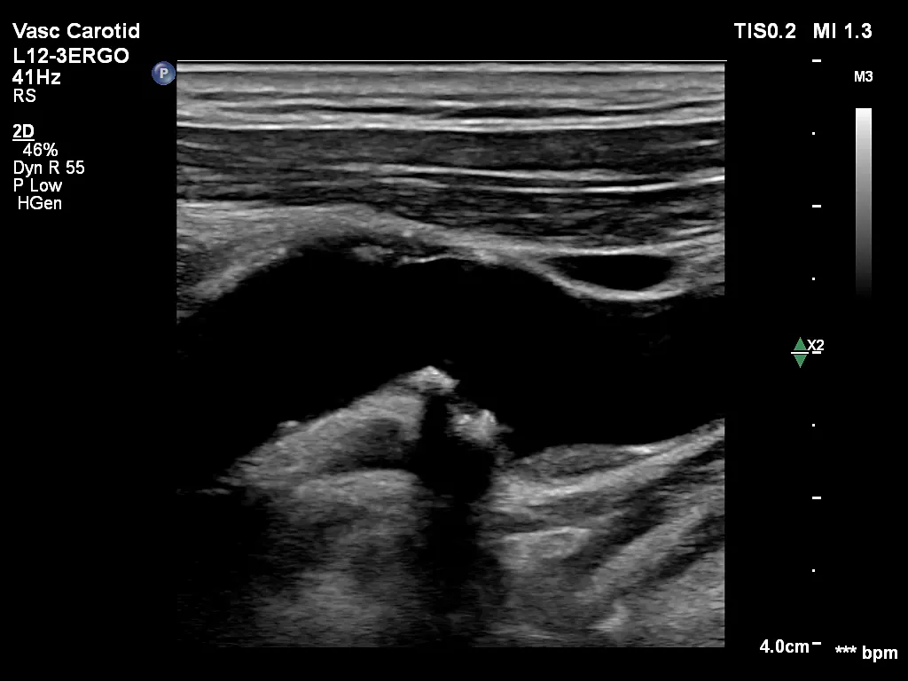
L12-3 paired with CEUS Auto Scan, scanning on the Vascular Carotid Preset
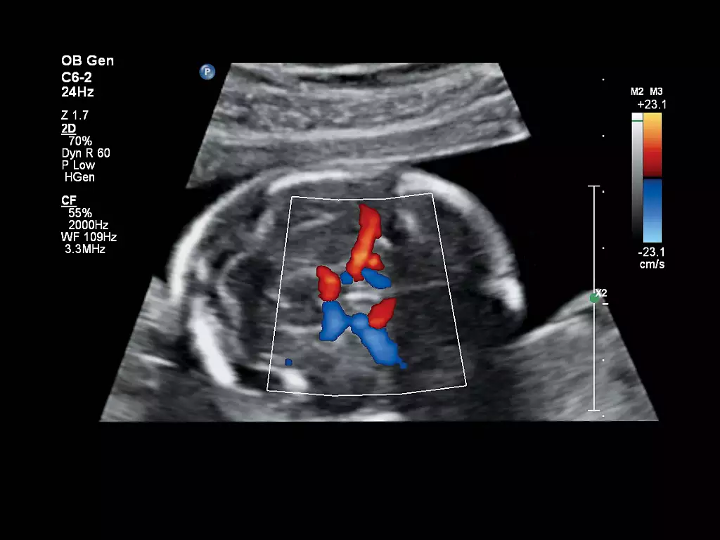
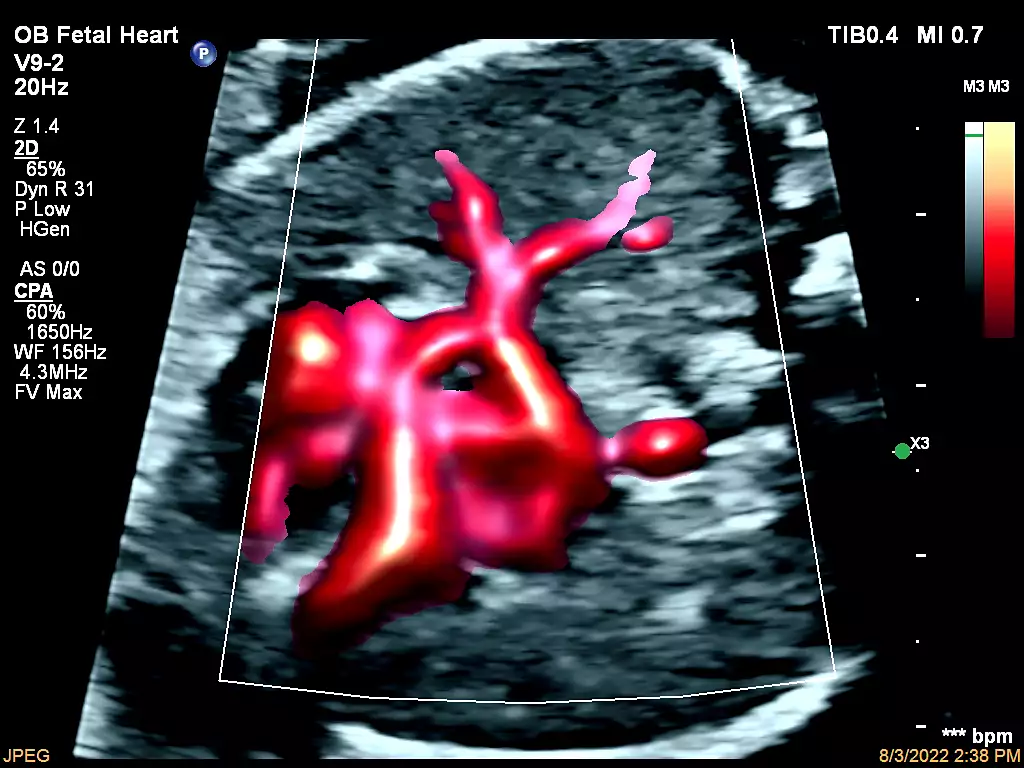
C6-2 OB Imaging V9-2 Fetal Heart CPA with Flow Viewer

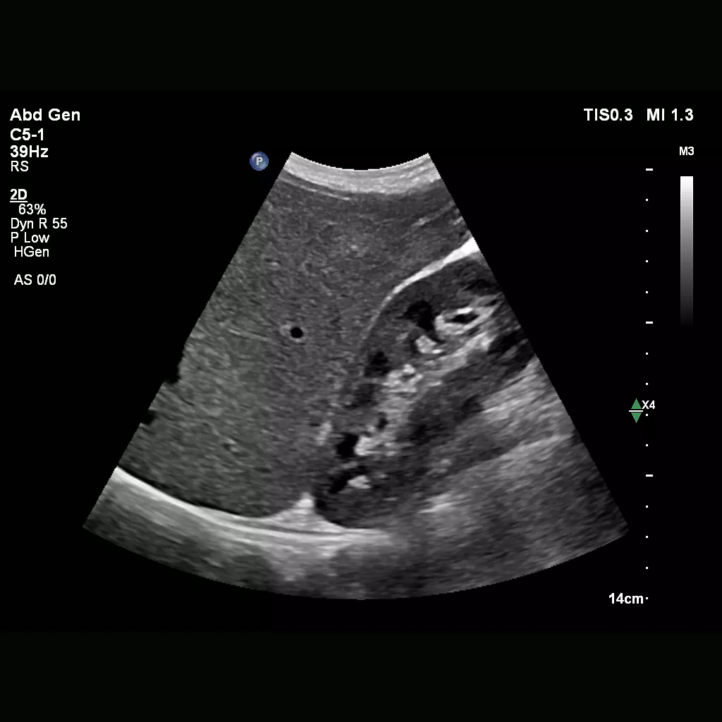
C5-1 Abdomen Liver
.webp)
L12-3ERGO Lower Extremity Calf Vessels

mC12-3 Neonatal Head C5-1 Pancreas Imaging
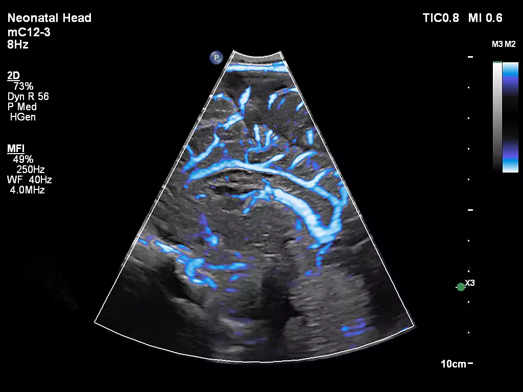
mC12-3 Neonatal Head MFI
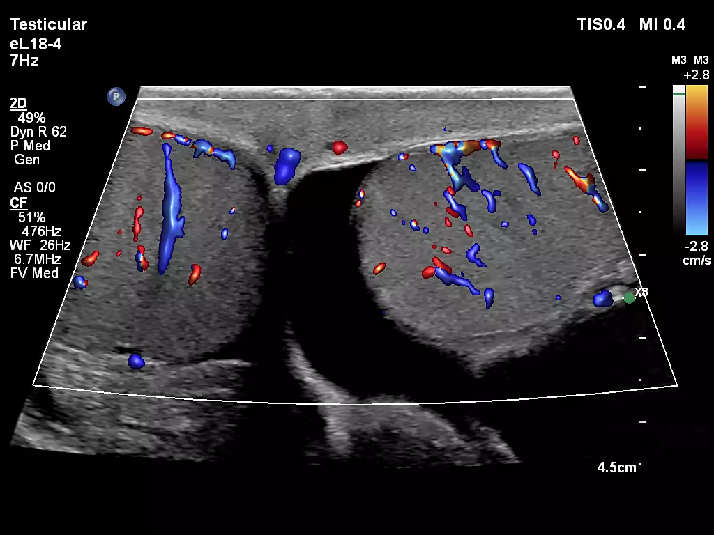
eL18-4 Testicular Imaging with FlowViewer
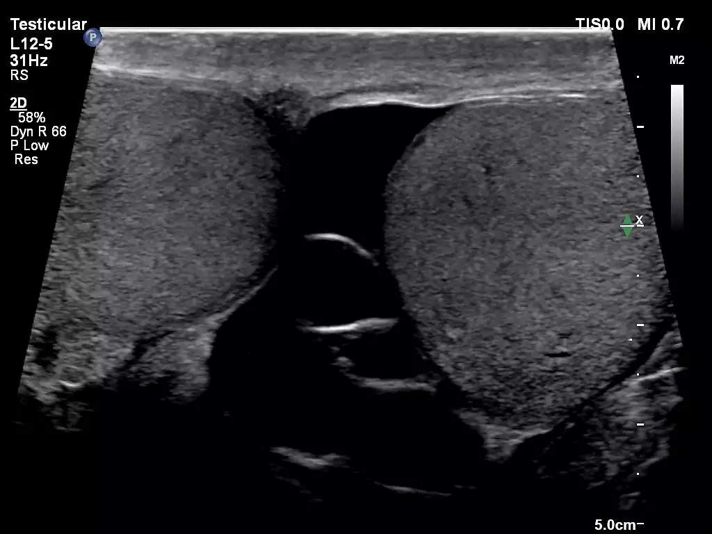
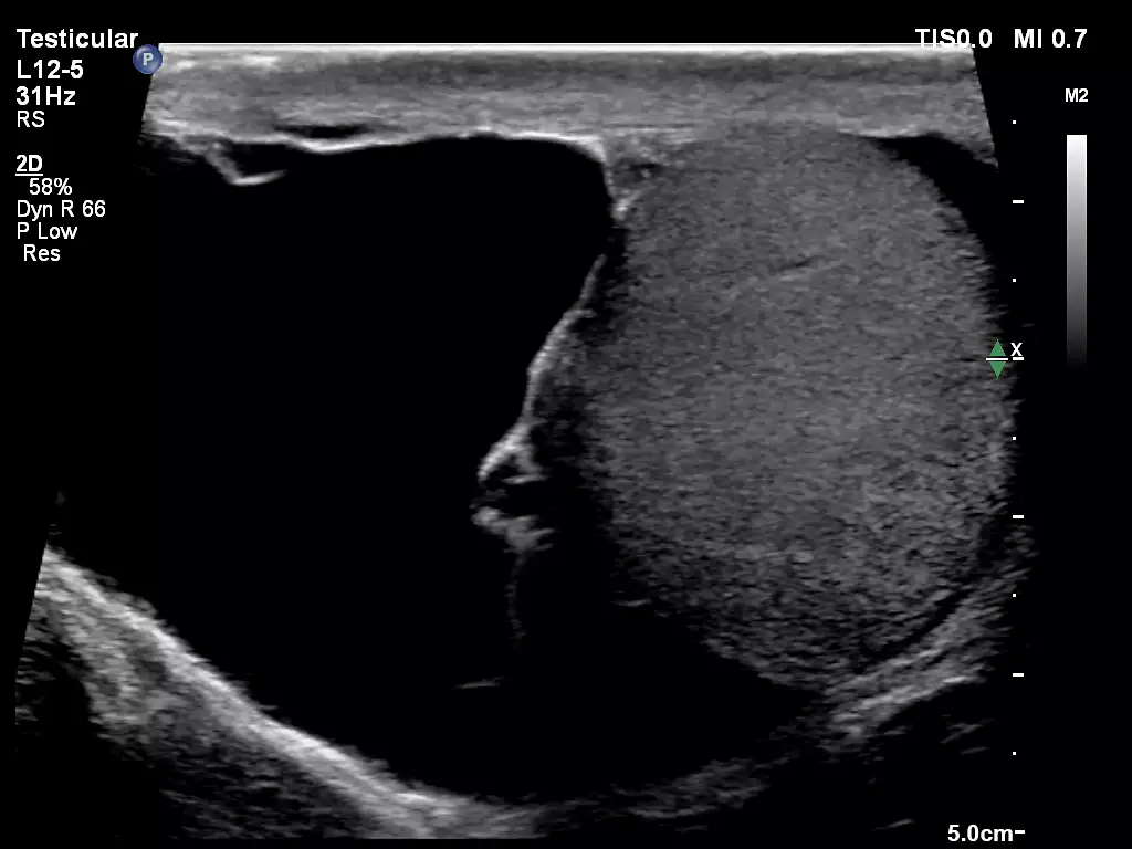
L12-5 Testicular Imaging L12-5 Testicular Imaging
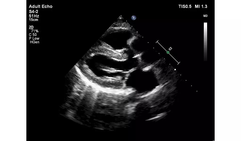
C5-1 Liver S4-2 PLAX showing Ventricular Septal Defect


 Microsystem
 Endoscopysystem
 Energysystem
 EndoscopyConsumables
 +86-21-54286005
 Room 602, Building 1, No. 111 Luxiang Road (Greenland Park Plaza), Baoshan District, Shanghai, China
  English
 中文
中文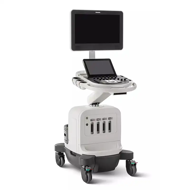
 Affiniti 50_Specificationen_EN

 中文
中文 English