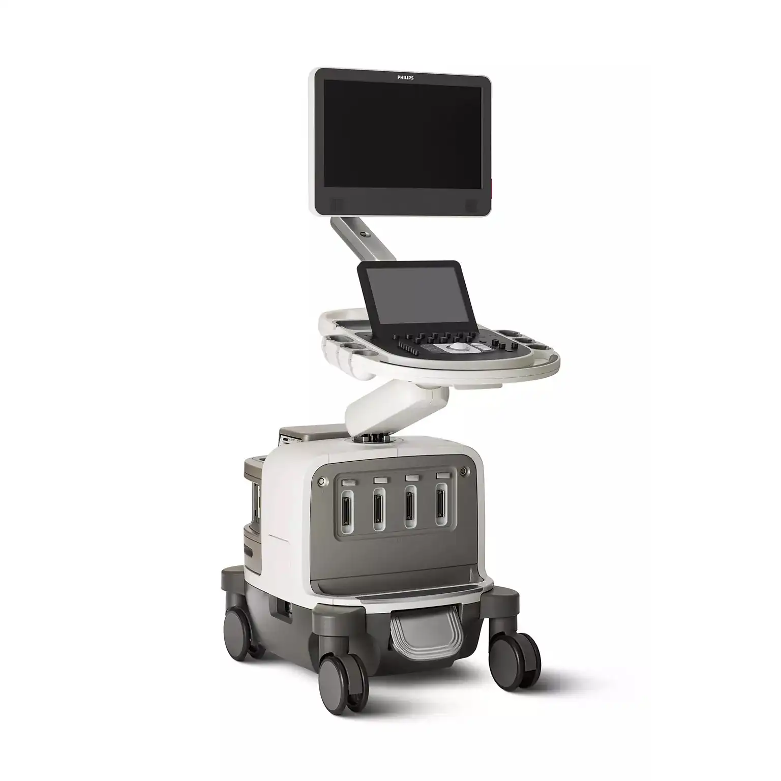
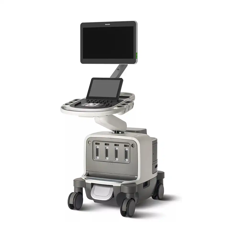
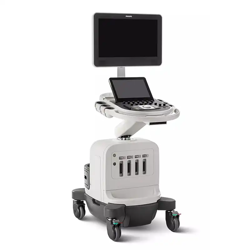
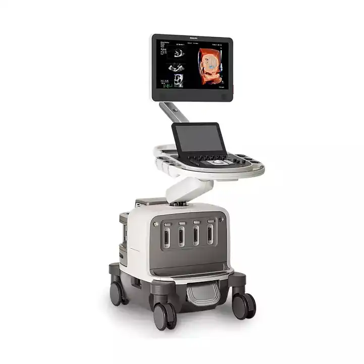
PHILIPS Affiniti 70
Ultrasound system

Affiniti 70 Elevate is the most advanced system in the Affiniti family, delivering stunning image quality and a suite of premium clinical features. Offering new levels of diagnostic confidence and reproducibility, it is designed for fast-paced environments with enhanced workflow and robust performance—helping you deliver the best possible care every day.
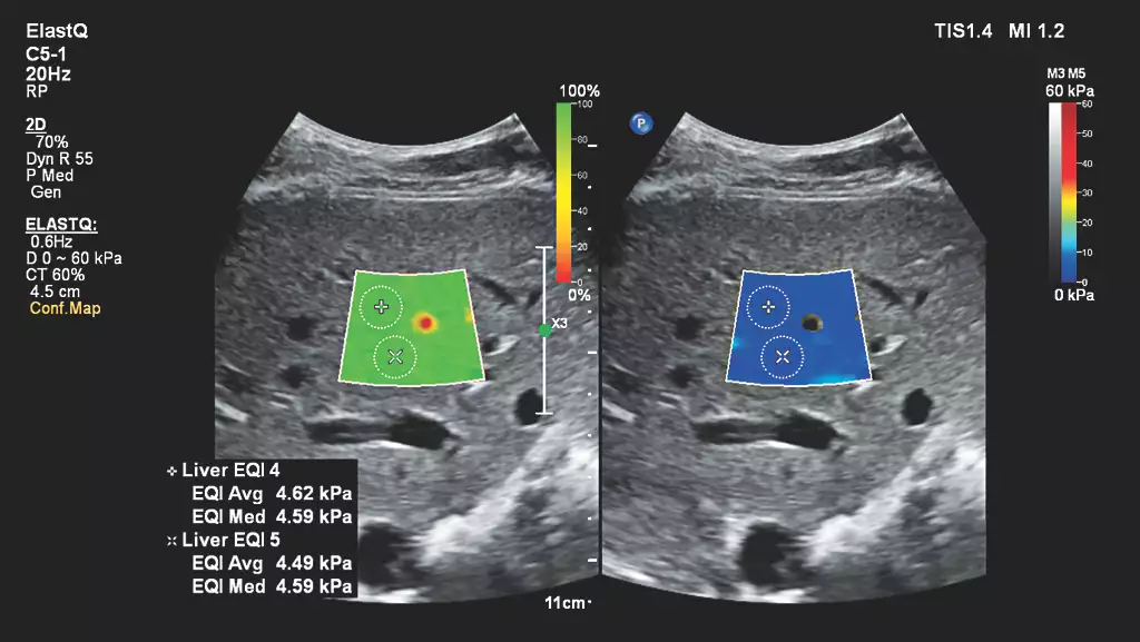
Auto ElastQ
Perform automated liver elastography with Auto ElastQ, and experience our next generation of liver health assessment. Auto ElastQ is designed to simplify user workflow with real-time, quantitative shear wave measurements.
Contrast-enhanced ultrasound (CEUS)
CEUS can transform the role of ultrasound in the liver, allowing the study of the enhancement patterns of suspicious liver lesions in real time, as well as provide an alternative non-ionizing approach to the assessment of vesicoureteral reflux in pediatric patients.
_EN.webp)
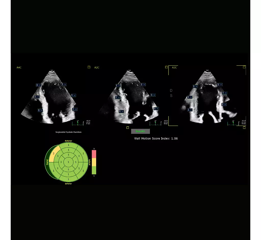
Auto Segmental Wall Motion Scoring
Provides automated evaluation of wall motion in a standard 17-segment bullseye display to aid objective LV wall assessment. With Auto SWMS, you can achieve greater reproducibility and efficiency in your workflows.
Next Gen Auto Scan
Philips Next Gen Auto Scan improves image uniformity, adaptively adjusting image brightness at every pixel and reducing the need for user adjustment while also improving transducer plunkability. Next Gen Auto Scan reduces button pushes by up to 54% with pixel-by-pixel real-time optimization.
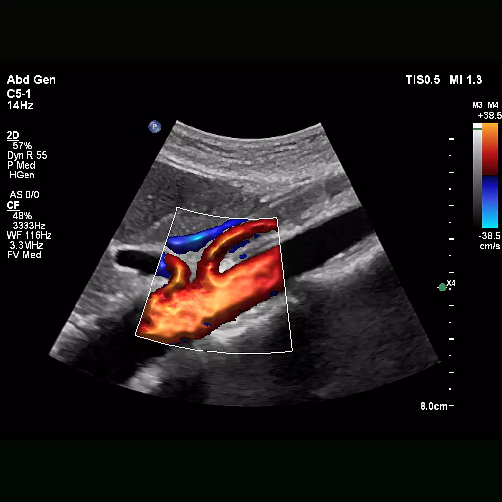
Flow Viewer
Flow Viewer is a Philips color visualization enhancement for vasculature and fetal heart architecture. Flow Viewer provides a 3D-like rendering of flow imaging data to better visualize the cardiac and vascular architecture and enhance the aesthetic appeal of all color imaging modes Available in all color imaging modes (CFM, CPA, CPAd, MFI, MFI HD).
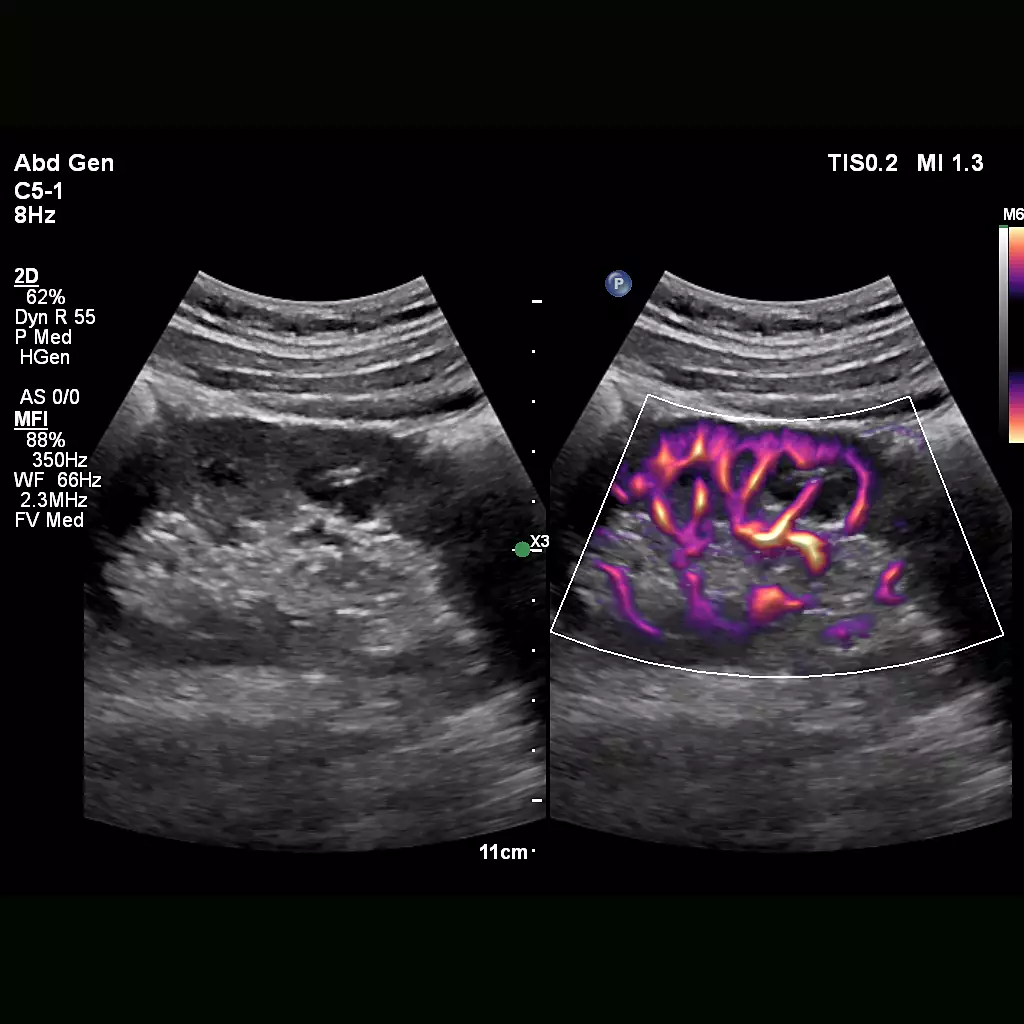
MFI
Designed to detect slow and weak blood flow anatomy in tissue. This proprietary approach overcomes many of the barriers associated with conventional methods to detect small vessel blood flow with high resolution and minimal artifacts. MicroFlow Imaging maintains high frame rate and 2D image quality while applying advanced artifact reduction techniques to reveal small vessel anatomy
Collaboration Live
Extend your team without expanding it. Collaboration Live is a communication platform that facilitates communication between a compatible ultrasound system and a remote user. With simultaneous Multi-party communication up to six users can quickly and securely talk, text, screen share and video stream directly from the ultrasound system for access to multiple clinical resources at a distance.

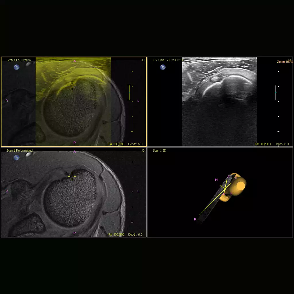
Fusion and Navigation
Make confident decisions even in challenging diagnostic cases with fully integrated fusion capabilities that feature streamlined workflows to allow clinicians to achieve fast and effective fusion of CT/MR/PET with live ultrasound. By combining imaging modalities directly on the ultrasound system, you now have access to an even more powerful diagnostic tool with advanced visualization allowing for fast clinical decisions. Expand fusion and navigation capabilities through a range of transducers across applications, including the X6-1 xMatrix , C5-1, C9-2, eL18-4, L12-5, C10-4ec, S5-1 and the new mC7-2
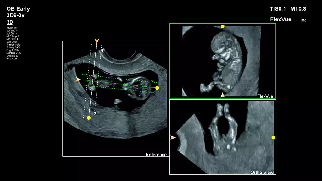
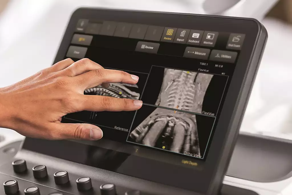
FlexVue with Orthogonal View
Easy-to-use tools designed to extract challenging anatomical planes from 3D data sets. This advanced feature offers exceptional flexibility in plane acquisition, complemented by a comprehensive measurement package for precise quantification.
PureWave & xMatrix transducer technology
The power of PureWave for exceptional imaging even on technically difficult patients. PureWave crystal technology represents the biggest breakthrough in piezoelectric transducer material in 40 years. The pure, uniform crystals of PureWave have virtually perfect uniformity for greater bandwidth and twice the efficiency of conventional ceramic materials.
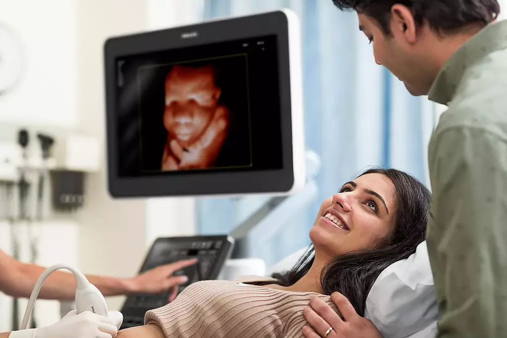
TrueVue advanced 3D display
Philips TrueVue advanced 3D ultrasound display delivers amazing lifelike 3D images. TrueVue, with its internal light source gives clinicians the ability to manipulate light and shadow anywhere in the 3D volume.
MaxVue high definition display
At the touch of a button, MaxVue high-definition display brings extraordinary visualization of anatomy with 1,179,648 additional image pixels compared to a standard 4:3 display format mode. MaxVue enhances ultrasound viewing and provides 38% more viewing area to optimize the display of dual, side/side, biplane, and scrolling imaging modes.
It understands your everyday
Philips Affiniti delivers the right balance of advanced ergonomic design and precision engineering to help you work more comfortably and intuitively. Its exceptional image quality gives you the results you need to provide the best patient care possible. See what Philips Affiniti can do for your practice.
Clinical image gallery
C5-1 Liver Fat Quantification eL18-4 MSK Shoulder
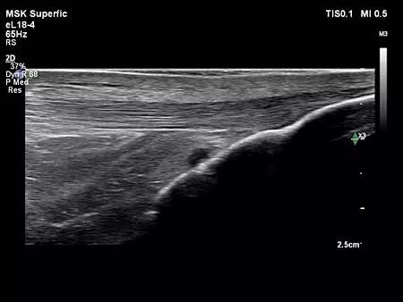
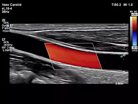
eL18-4 MSK Shoulder eL18-4 Vascular Carotid

eL18-4 Adv Breast
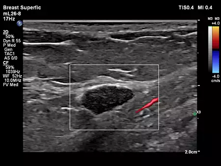
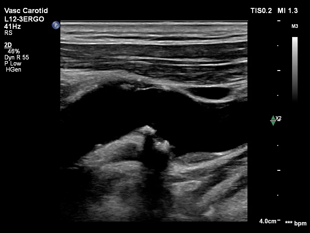
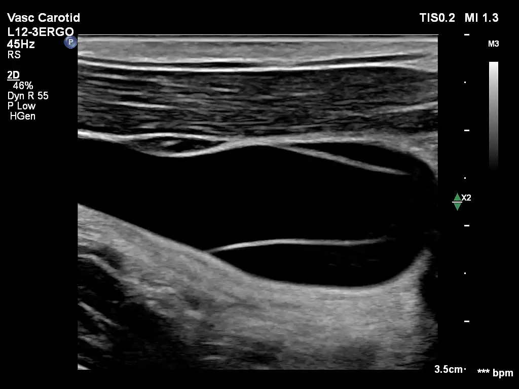
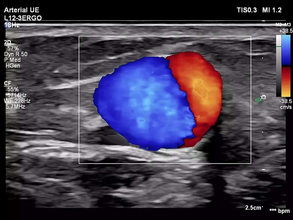
mL26-8 Breast L12-3ERGO Vascular Carotid Bulb
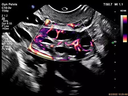
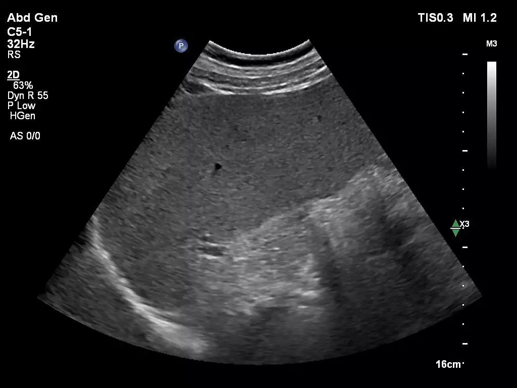
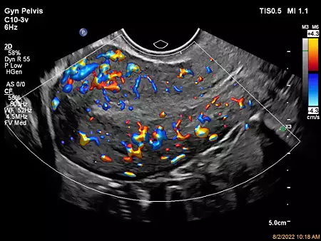
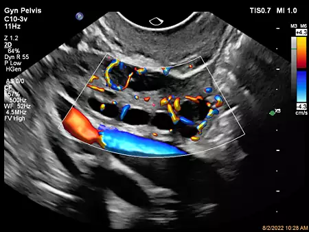
C10-3v Ovary with MicoFlow Imaging C5-1 Abdomen Spleen Imaging

eL18-4 MSK Patellar Tendon eL18-4 OB Imaging
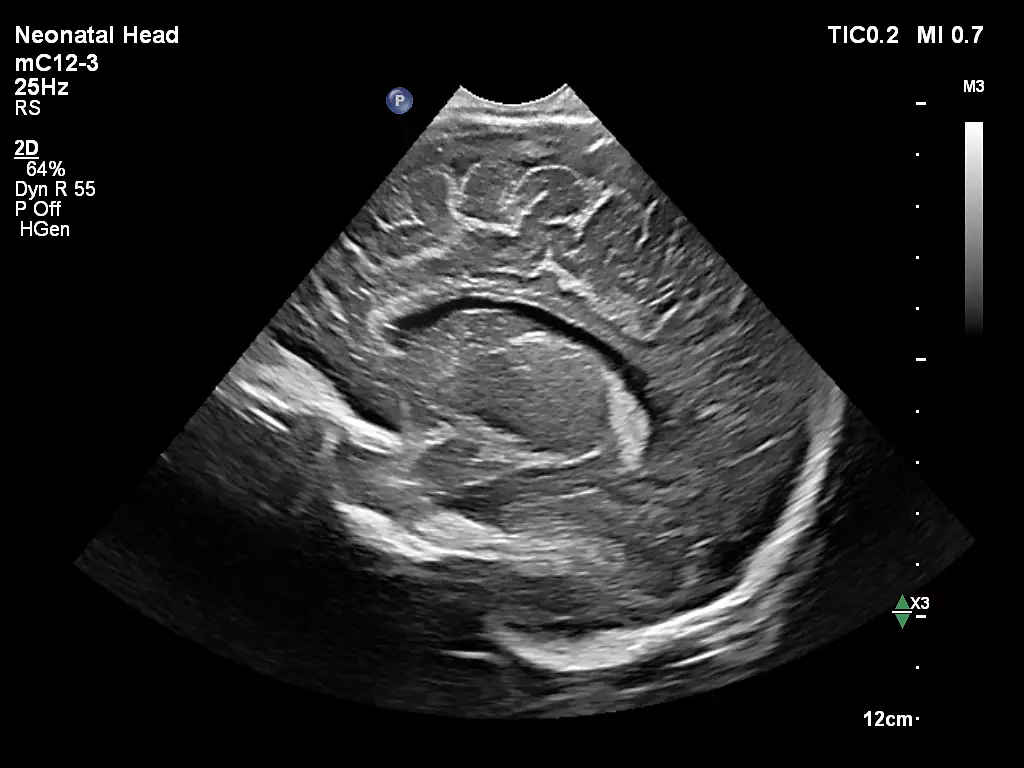
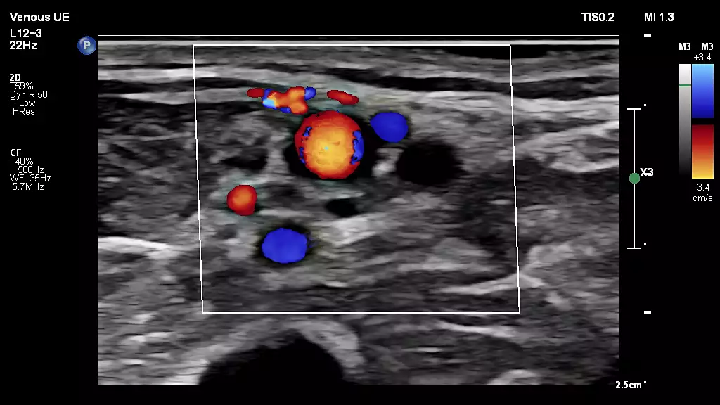
L12-3ERGO Internal Jugular Vein mC12-3 Neonatal Head
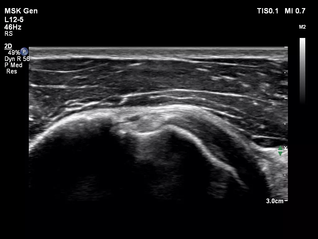
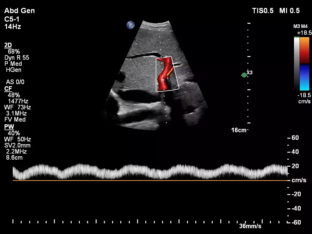
L12-5 MSK Biceps Tendon C5-1 Abdominal Proximal Aorta with Flow Viewer
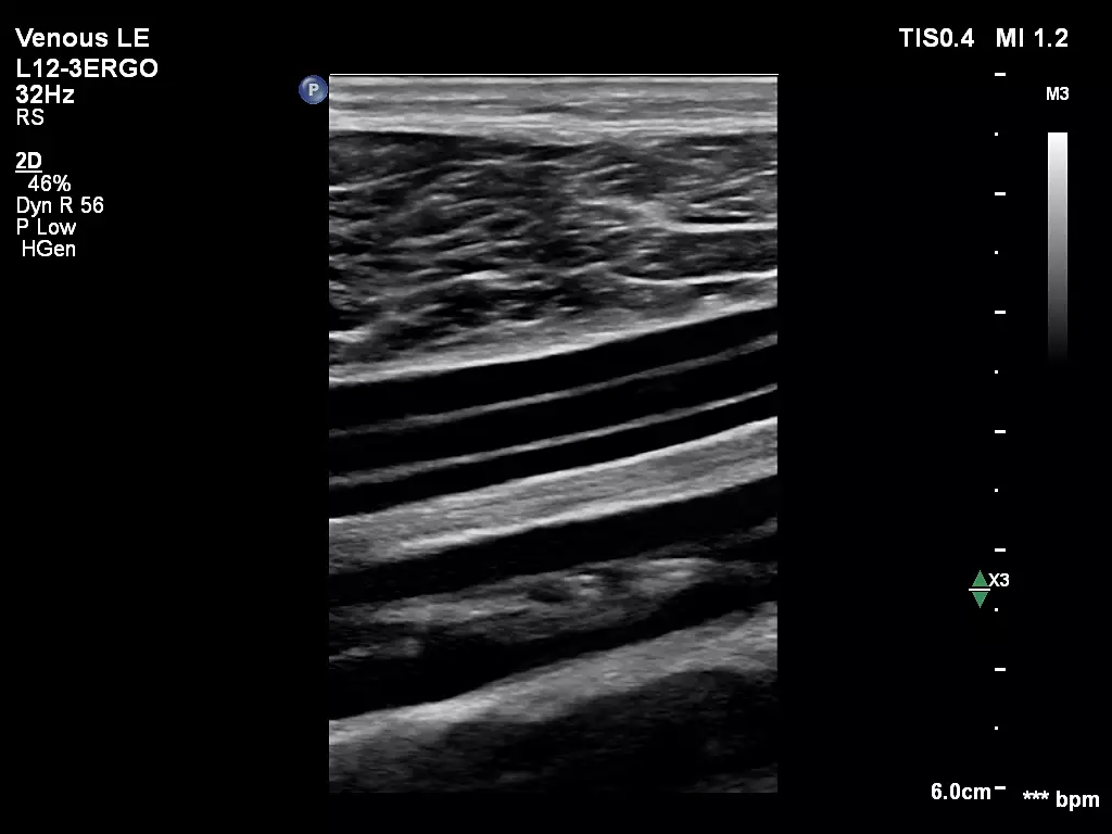
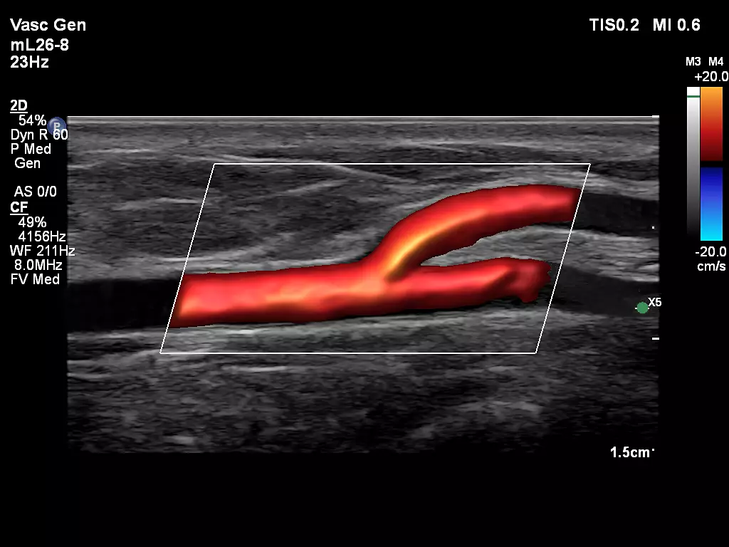
L12-3ERGO Lower Limb Veins mL26-8 Superficial Artery with FlowViewer
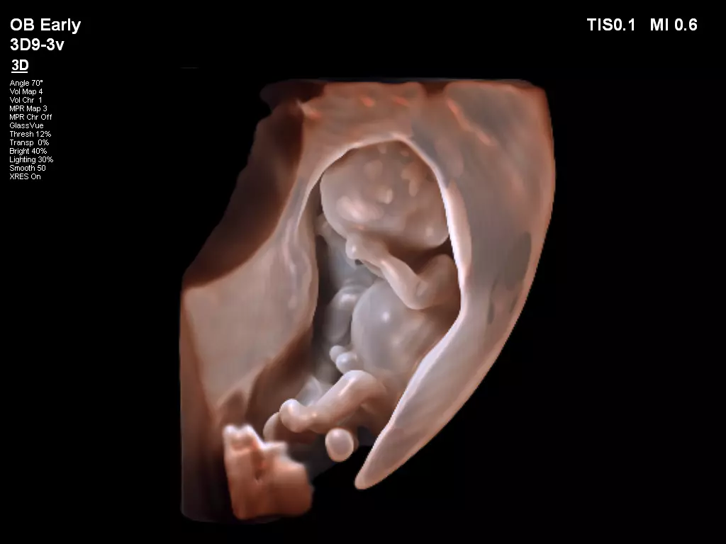
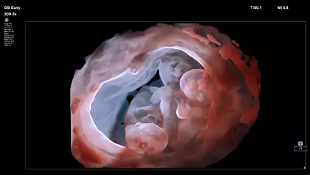
3D9-3v Fetal Imaging with TrueVue L12-3ERGO Fistula Imaging
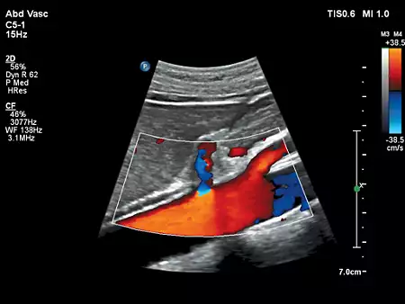
C5-1 Abdominal Vascular C5-1 Liver Doppler with FlowViewer
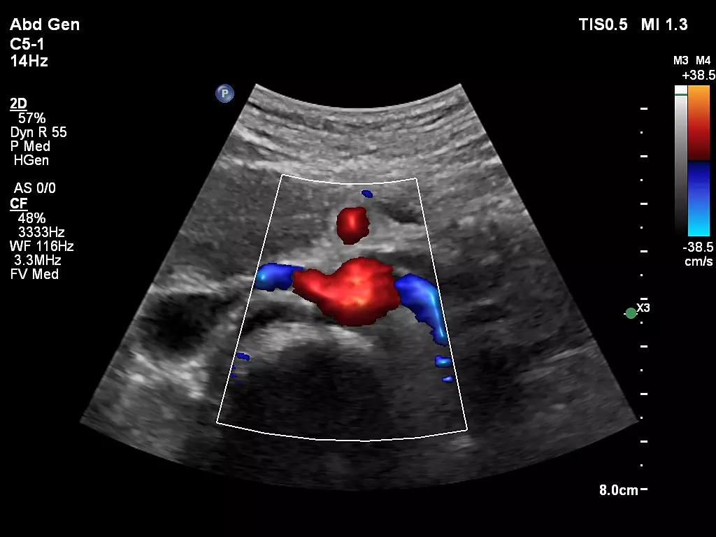
C5-1 Renal Artery Origin with FlowViewer 3D9-3v Fetal Imaging with GlassVue
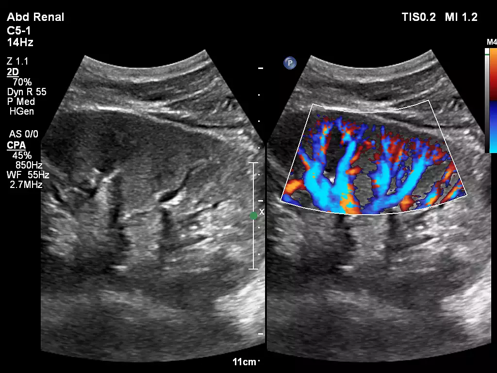
C5-1 Renal Kidney Imaging C10-3v Ovarian Perfusion with Flow Viewer
_EN.webp)
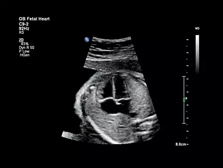
C5-1 Hepatic Renal Index (HRI) C9-2 Fetal heart
eL18-4 Testicular Imaging with MicroFlow Imaging


 Microsystem
 Endoscopysystem
 Energysystem
 EndoscopyConsumables
 +86-21-54286005
 Room 602, Building 1, No. 111 Luxiang Road (Greenland Park Plaza), Baoshan District, Shanghai, China
  English
 中文
中文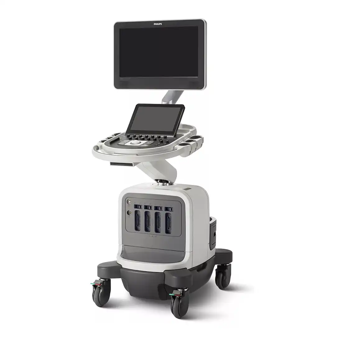
 Affiniti 70_Brochure_EN

 中文
中文 English