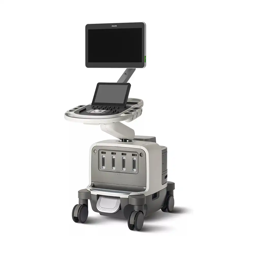
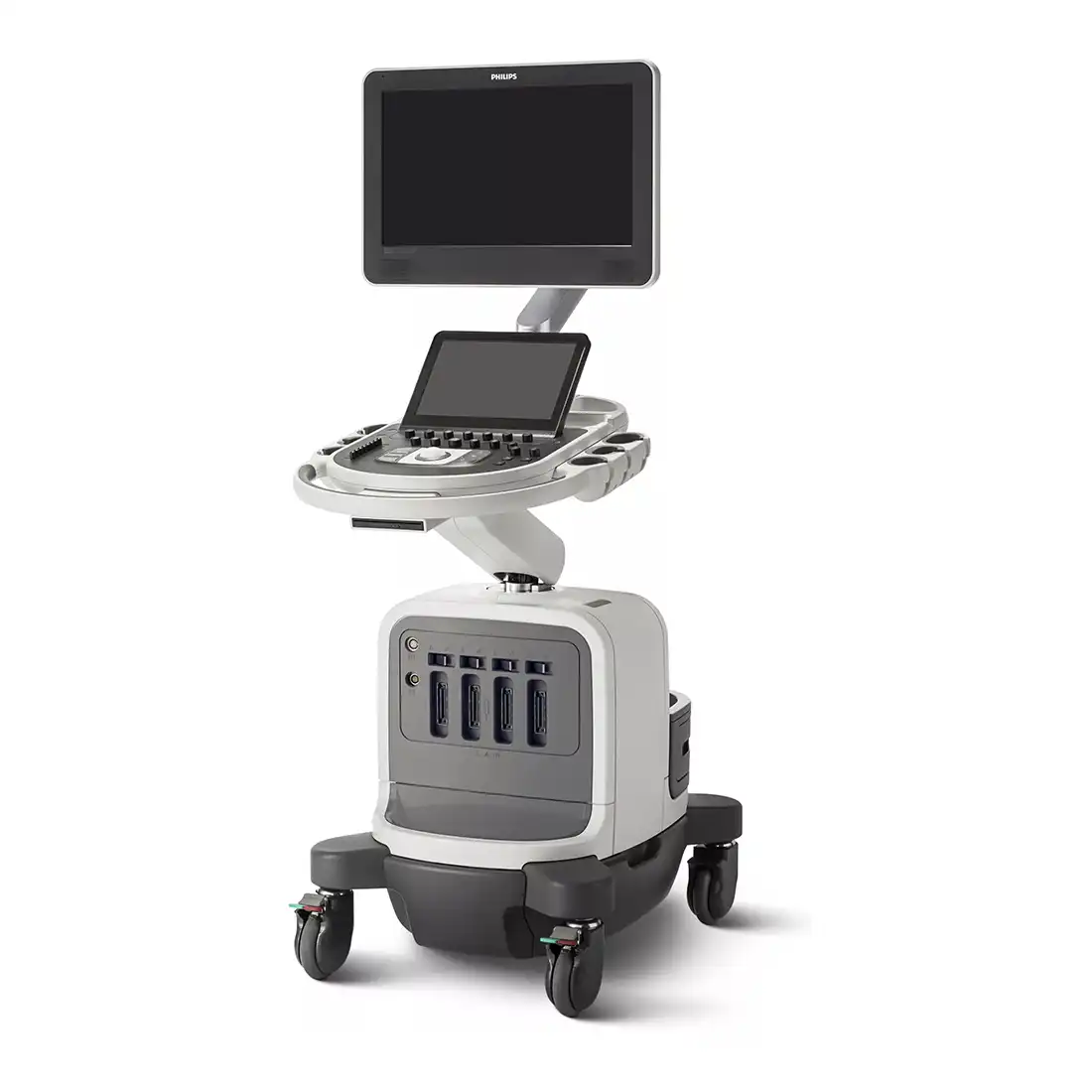
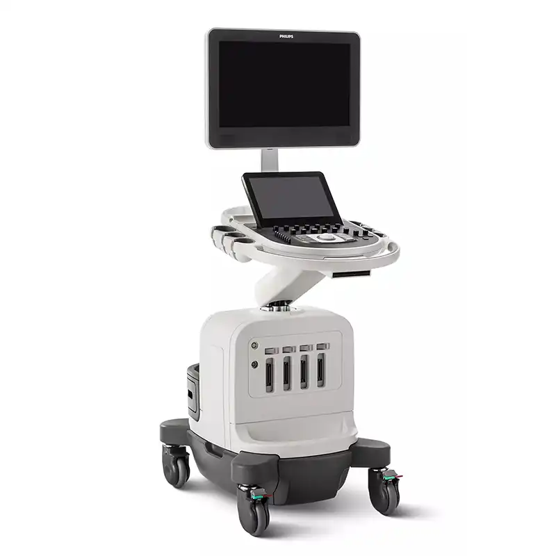
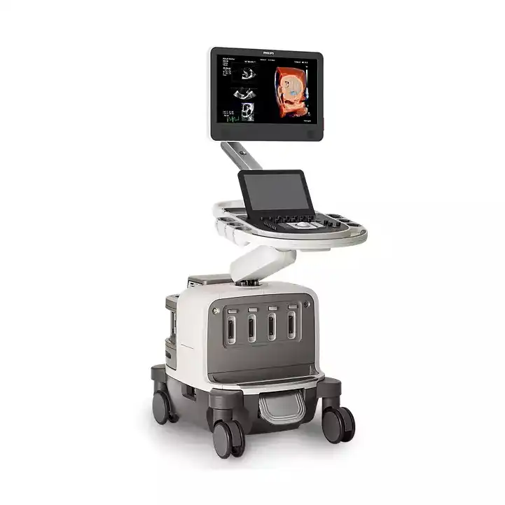
PHILIPS EPIQ CVx,CVxi
Premium Cardiology Ultrasound System

EPIQ CVx,CVxi, our premium cardiovascular ultrasound system built on our innovative, modular, industry-leading ultrasound platform, has powerful AI-based capabilities and advanced diagnostic solutions to help you transcend today's complexities and propel echocardiography into the next dimension. This enables you to achieve greater consistency, accessible innovation, smarter workflows, and easier scalability.
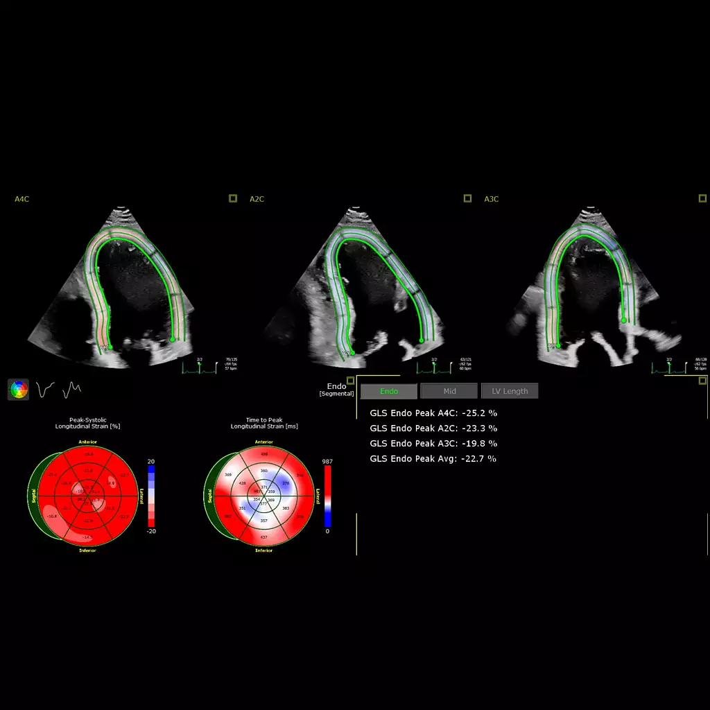
AutoStrain LV with automated EF and mid-layer strain
AutoStrain delivers fast, reproducible 2D strain quantification for the LV, LA, and RV, as well as automated EF and mid-layer strain for a comprehensive LV assessment within the same application, improving workflow and saving time. Smart View Select works in the background and uses AI to automatically select the optimum images for 2D LV assessment.
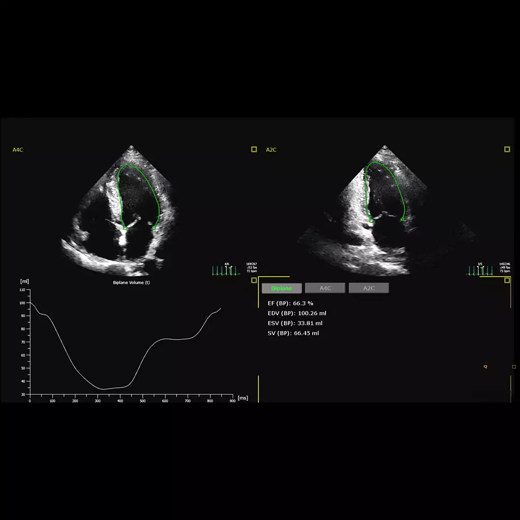
2D Auto EF and 2D Auto EF Advanced
Offers fast, reproducible results for easy comparison of LV functional data to improve workflow and save time in LV assessments. 2D Auto EF supports non-contrast images, and 2D Auto EF Advanced uses AI to support the quantification of contrast and non-contrast images.
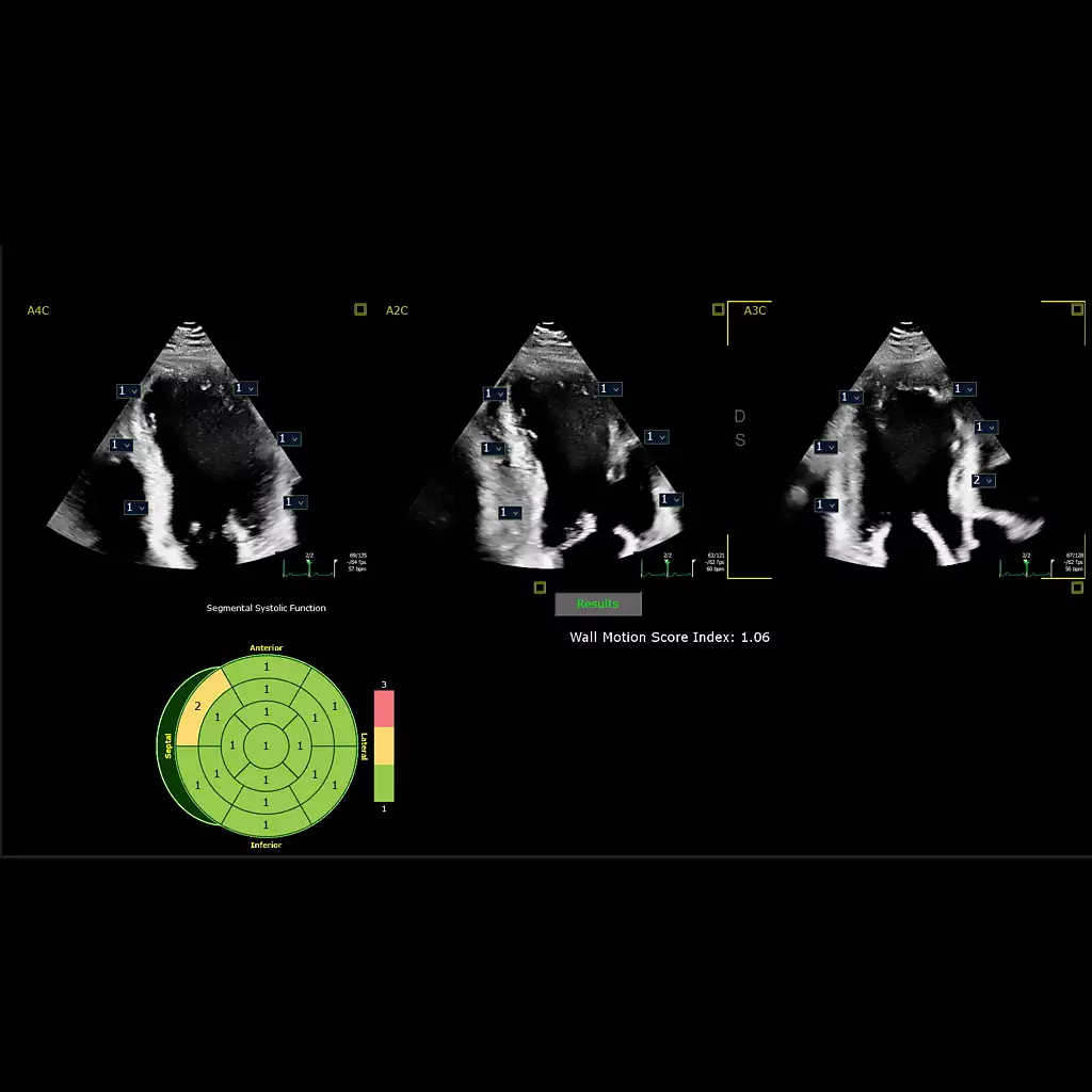
Auto Segmental Wall Motion Scoring
Provides automated evaluation of wall motion in a standard 17-segment bullseye display to aid objective LV wall assessment. With Auto SWMS, you can achieve greater reproducibility and efficiency in your workflows.
Auto Measure provides automation for robust, proven, reproducible cardiac quantification
Auto Measure provides fully automated 2D doppler and length measurements along with AI assisted Smart (Doppler) View ID which further enhances time-savings. AI provides full functionality without EKG.
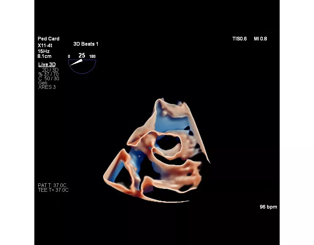
The Philips X11-4t mini TEE. It fits right.
When navigating narrow spaces and complex cases, the Philips X11-4t mini 3D TEE fits right, giving you the room, reach and angles to help diagnose and treat more patients. X11-4t is designed for both adults and pediatrics, down to 5kg. All delivered with the ease you know and the legacy you trust from Philips.
3D Markers
Graphic markers can be placed within a 3D Volume or MPR while in MultiVue to streamline tracking of structural points of interest. 3D Markers facilitate greater efficiency during echo-guided procedures and allow for detailed annotation of complex anatomy.
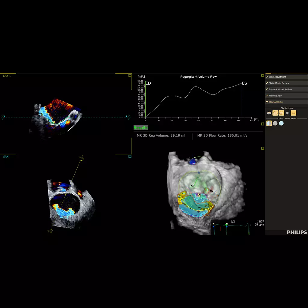
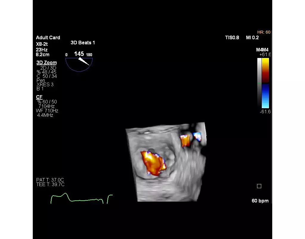
3D Auto Color Flow Quantification***
Offers AI for fast, easy and reproducible mitral valve regurgitation (MR) volume to help assess MR severity.
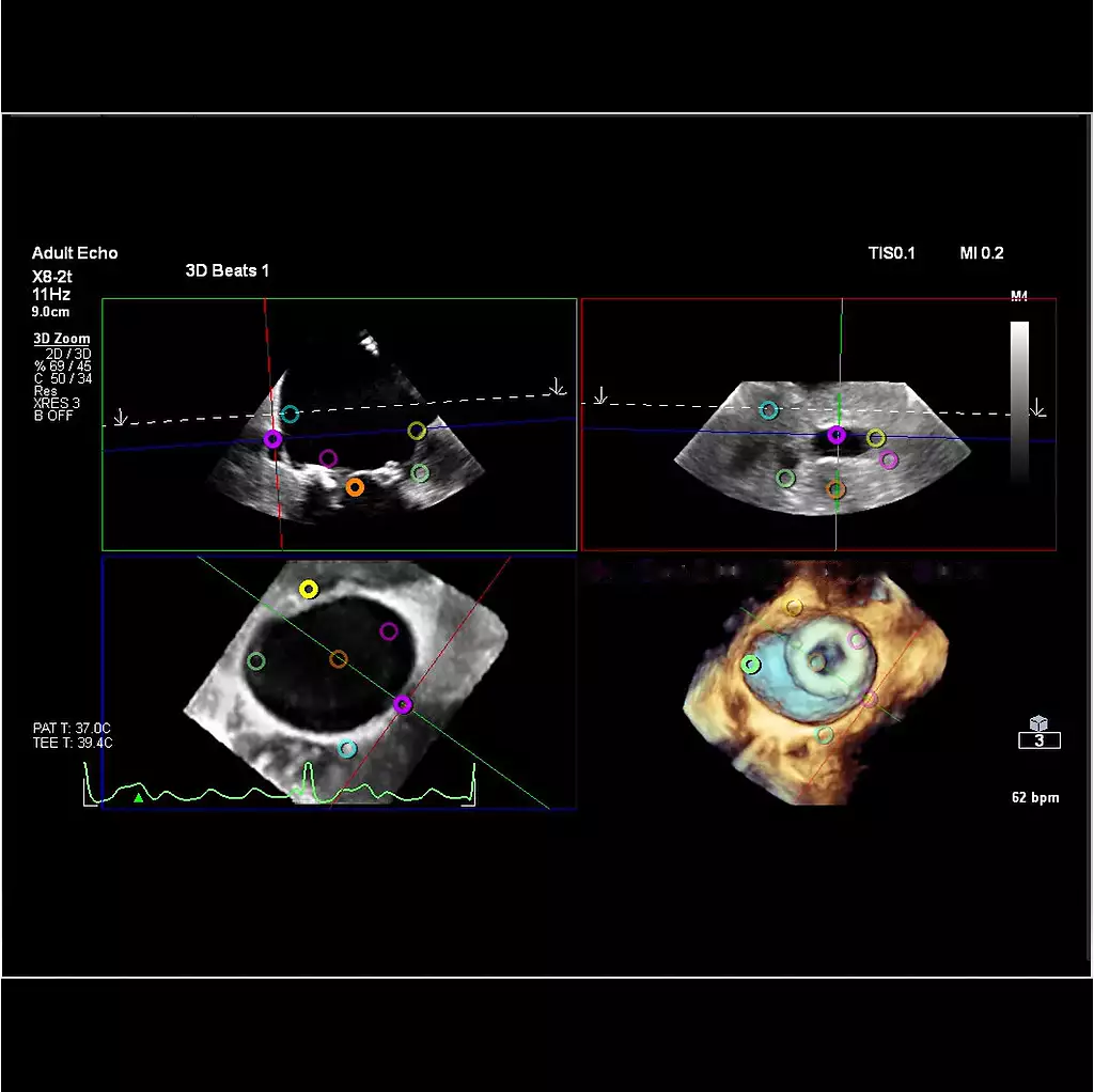
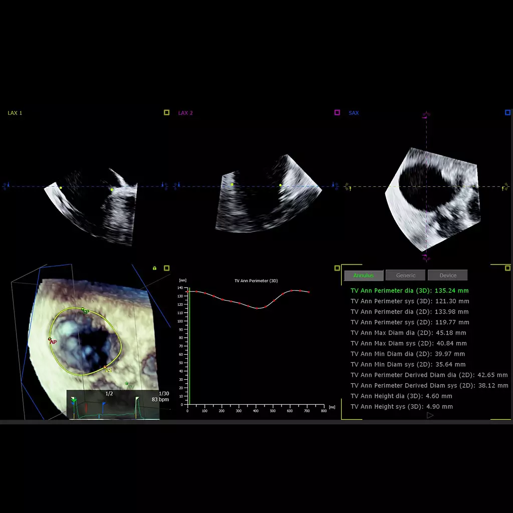
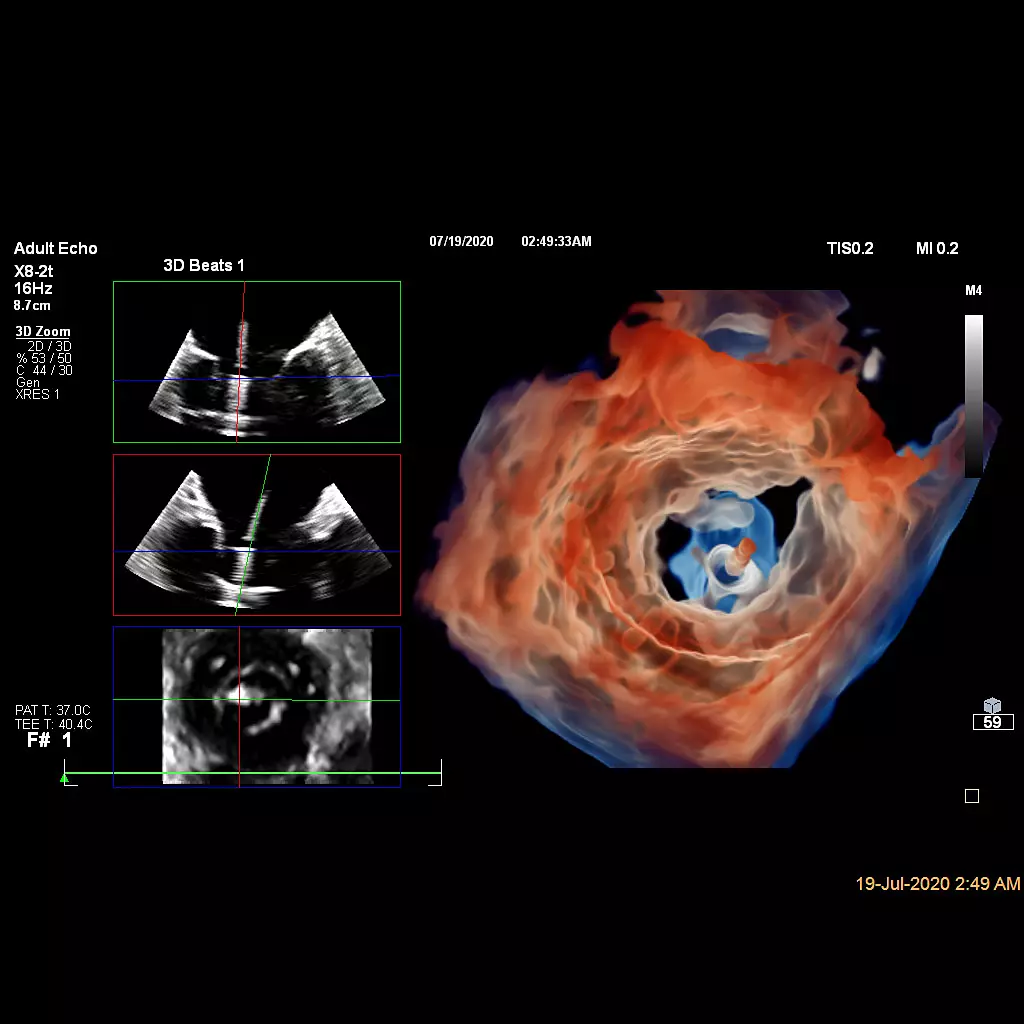
3D Auto Tricuspid Valve Quantification***
AI helps confirm/re-evaluate device size/selection with accurate and peri-procedure TV annulus measurements (initial sizing and plan with CT).
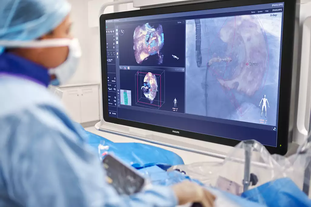
EchoNavigator
EchoNavigator automatically fuses live 3D TEE and live X-ray in real time. The solution assists heart teams with intuitive image guidance during procedures where both real-time X-ray and echo imaging are used and fused together. EchoNavigator provides automated fusion of echo and X-ray images, automated markers for context and guidance, and up to three different echo views simultaneously.
nSight Plus expansion to key transducers
X5-1c, X8-2t, and VeriSight Pro have the power of software beamforming. Now enhanced with nSight Plus, our transducers have improved MPR image quality and increased frame rates.
Efficiencies in TEE workflows
Efficient workflow for device positioning and size during procedures with MultiVue and Recall Settings. MultiVue provides one click alignment with 3D anatomy, where Recall Settings preserves acquisition and imaging settings when switching between ICE, TEE, and TTE transducers.
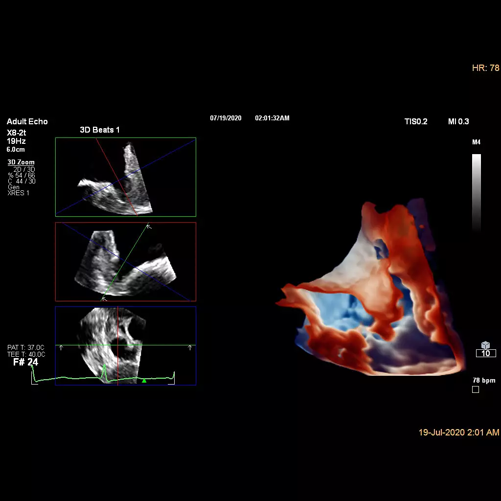
Ultrasound Left Atrial Appendage solution
Designed to provide real time LAA morphology from Live 3D imaging with Cardiac TrueVue Glass and to reduce time in LAA landing area quantification even during complex interventional procedures.


 Microsystem
 Endoscopysystem
 Energysystem
 EndoscopyConsumables
 +86-21-54286005
 Room 602, Building 1, No. 111 Luxiang Road (Greenland Park Plaza), Baoshan District, Shanghai, China
  English
 中文
中文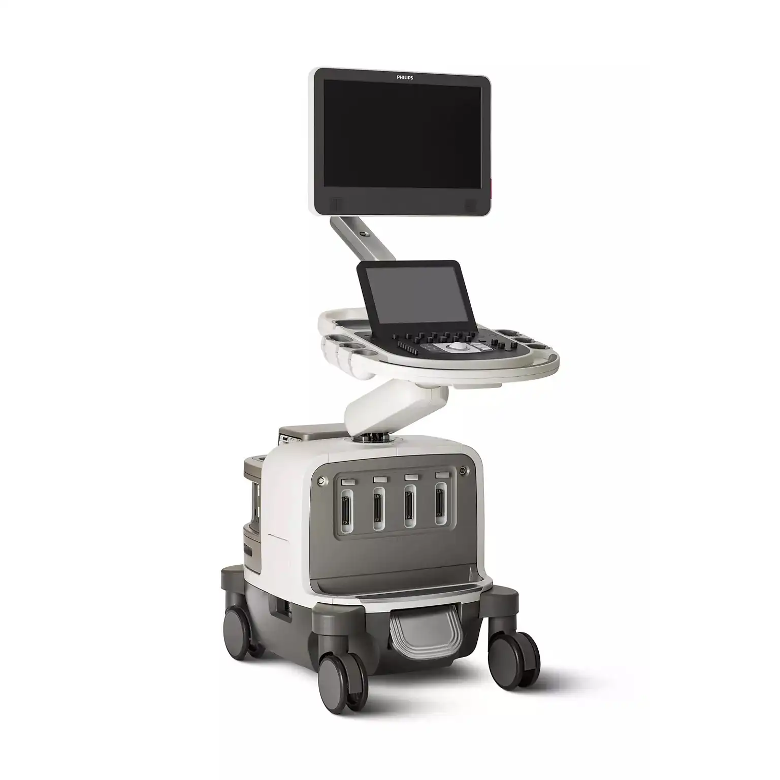
 PHILIPS EPIQ CVx,CVxi_Brochure_EN

 中文
中文 English