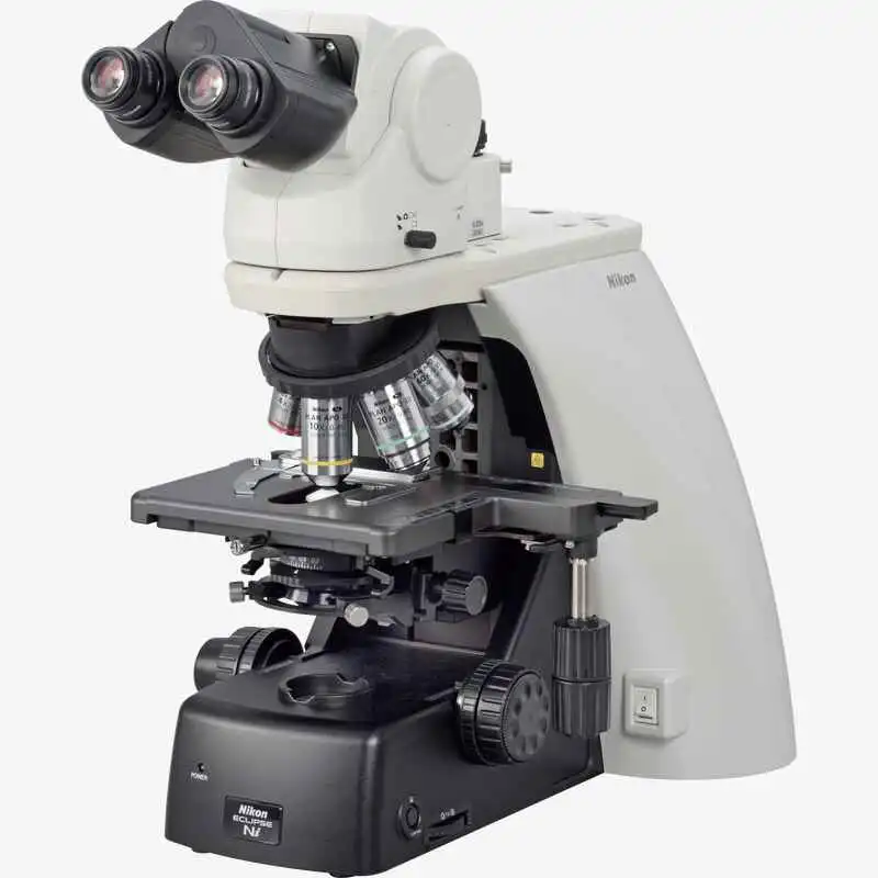
NIKON ECLIPSE Ci-E
Motorized Upright Biological Microscope
Clinical and laboratory microscopes featuring LED illumination, providing stress-free operation for routine use
To meet the demands of clinical laboratory specialists and researchers, Nikon has reviewed all aspects of microscope usability to develop the ECLIPSE Ci-E of microscopes, which combines superior functionality with operational ease. The Ci is designed to ensure natural posture while viewing images, sample changing and capturing images. Bright, eco-friendly LED illumination reduces the need for frequent lamp replacement. A variety of accessories are available that support various imaging techniques.

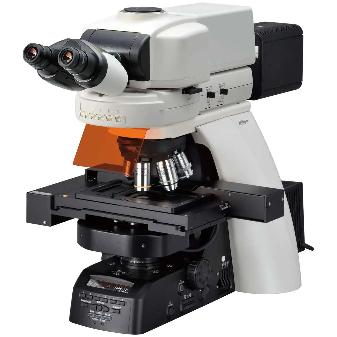
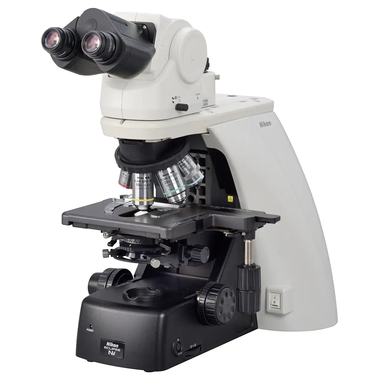
Motorized magnification switching
Magnification can be easily changed during observation with the touch of a button on the ECLIPSE Ci-E microscope body. The buttons can also be programmed for specific objective lenses for easy switching between specific pairs. User-defined light intensity for each magnification is automatically saved and reproduced when magnification is changed. The remote control pad can also be used to easily change magnifications.
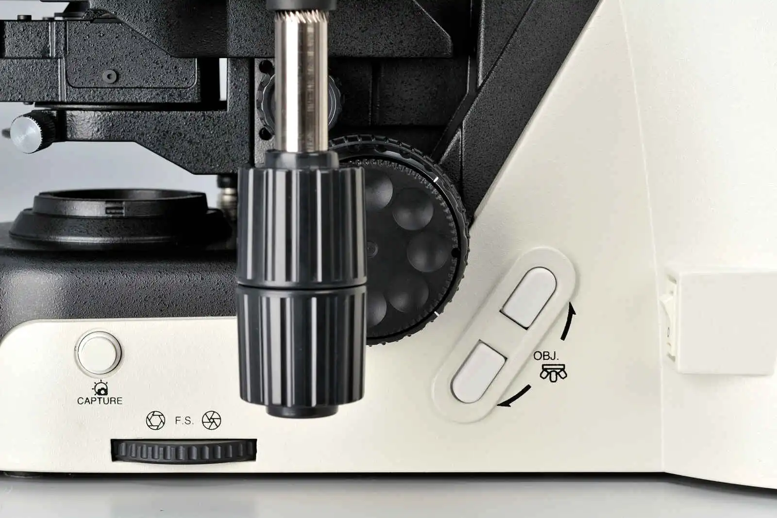
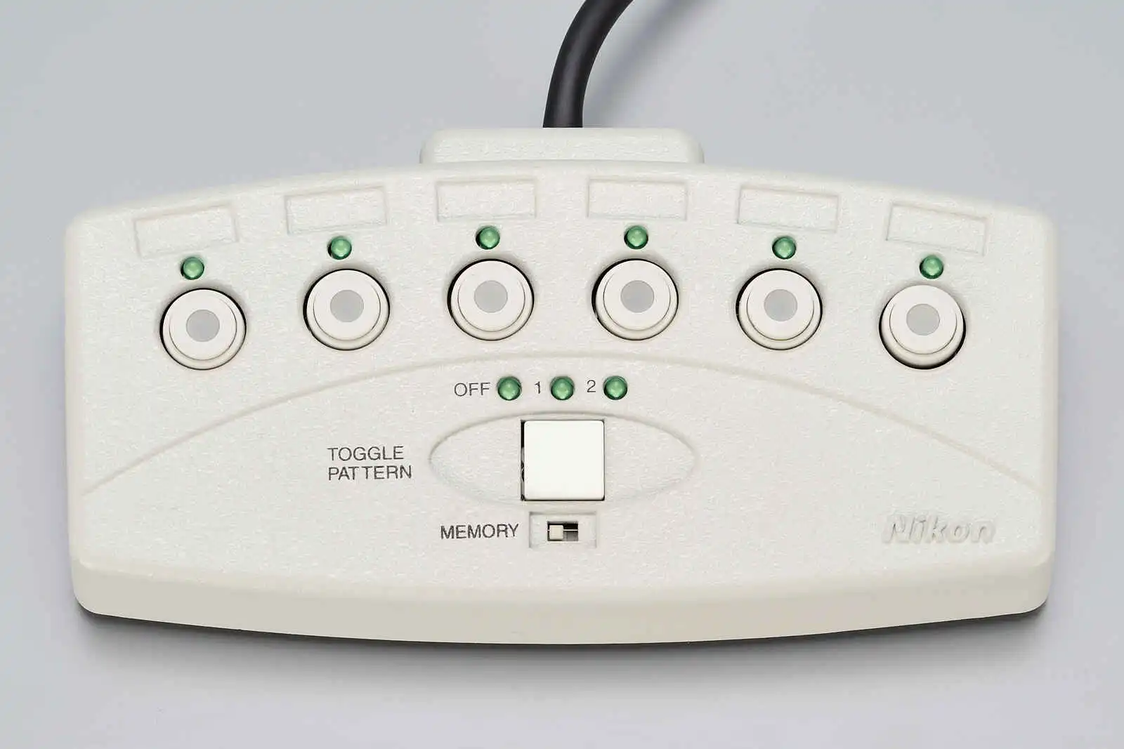
Nosepiece rotation buttons Remote control pad
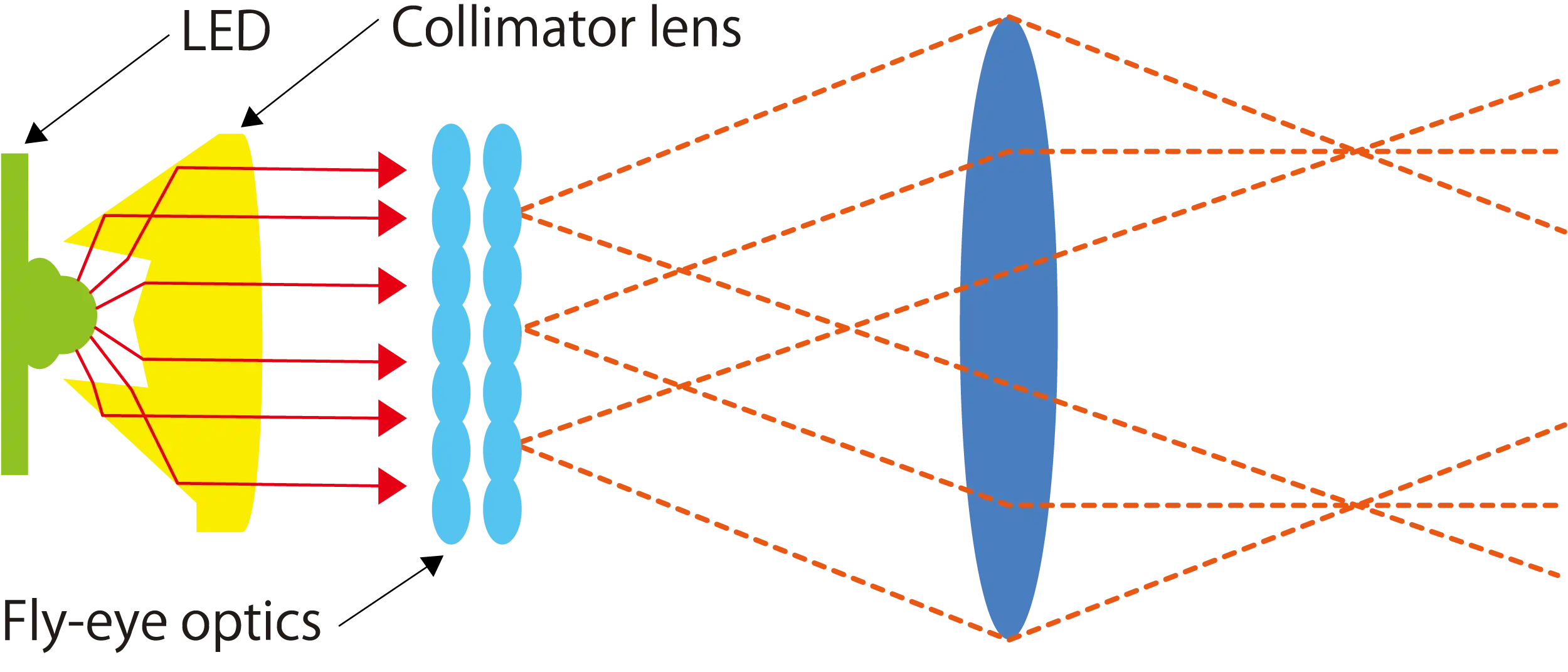
Bright and uniform Eco-illumination
The Eco-illumination is a low-power-consuming, eco-friendly illumination system that produces uniform brightness and reduces the cost and effort of lamp replacement, thanks to its 60,000 hour, high-luminescent LED. By combining a collimator lens, fly-eye optics and LED illumination, bright and edge-to-edge uniform images can be obtained even at high magnifications. The LED illuminator features low-heat generation and provides the same color temperature at every magnification.
Enhanced operational ease
Observation with a natural posture
Using the ergonomic binocular tube, which features an eyepiece that can be inclined from 10° to 30°and extended up to 40 mm, the microscope can be adjusted for natural posture. The eyelevel riser lifts the eyepiece tube in 25 mm increments (up to 100 mm*) and can easily be adapted to suit users with different eye-point heights.
* Up to 50 mm with ergonomic binocular tube.

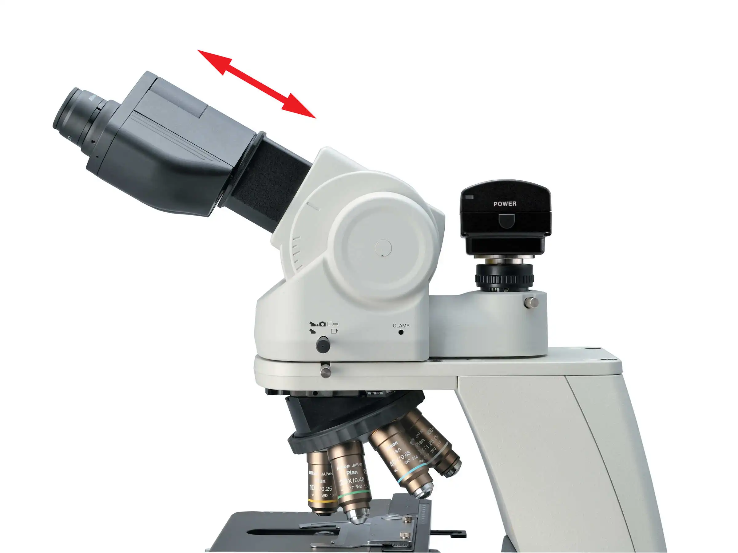
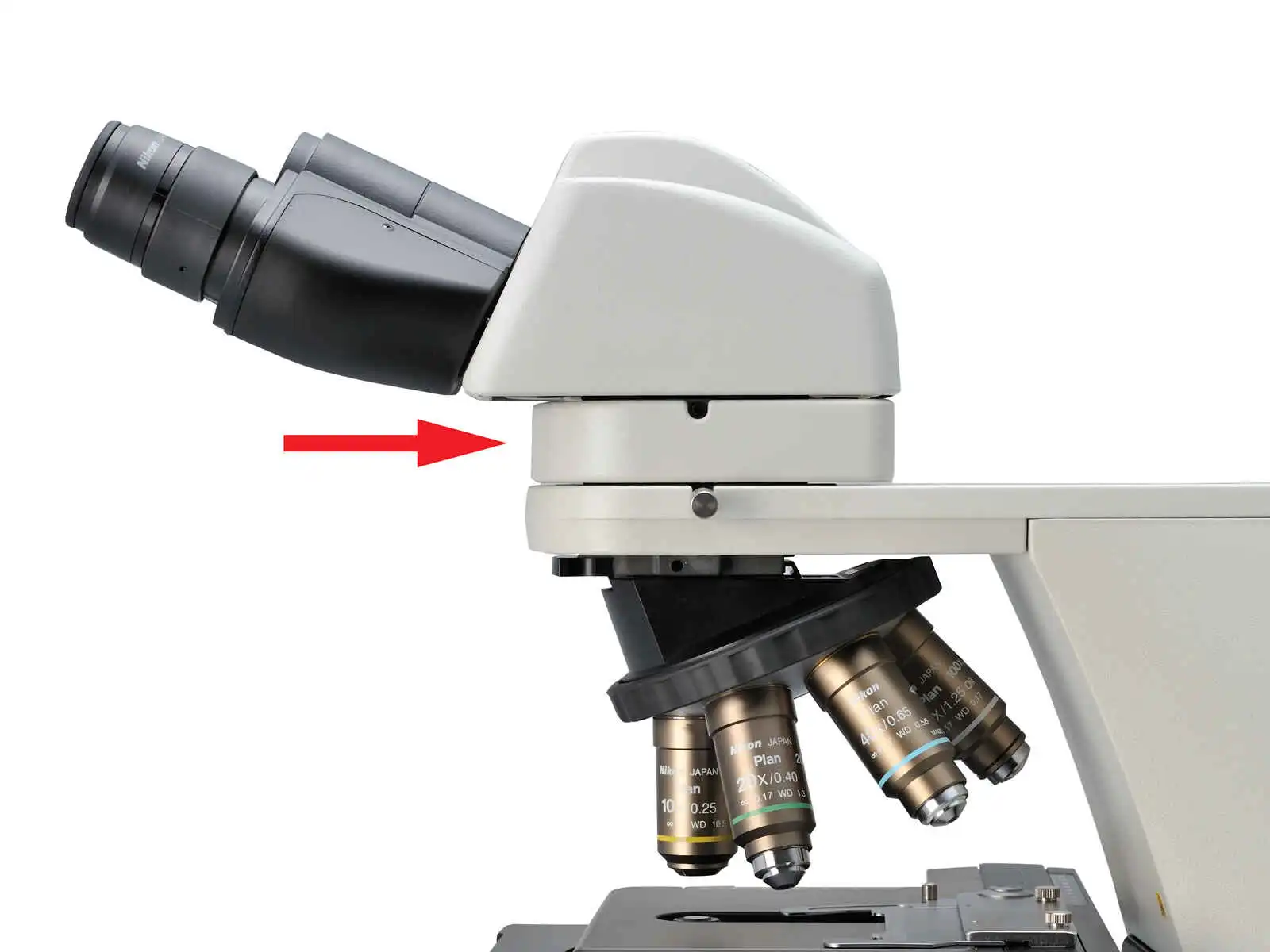
Ergonomic binocular tube Eyelevel riser
User-friendly stage operation
With the addition of a nosepiece spacer, the stage height can be lowered 20 mm from the standard position, reducing strain during frequent specimen change. The stage handle height can also be changed to ensure a comfortable hand position. The stage height can be locked using the refocusing knob, allowing quick refocusing after specimen changes. The stage is coated with a high-durability, scratch-resistant ceramic coating.
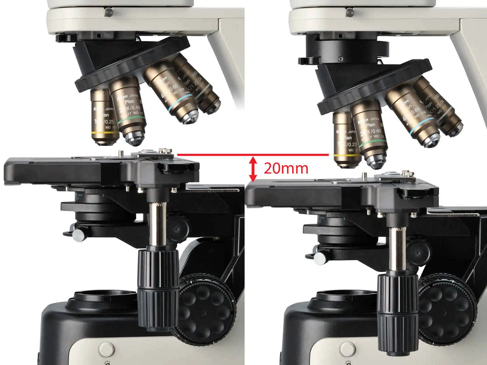
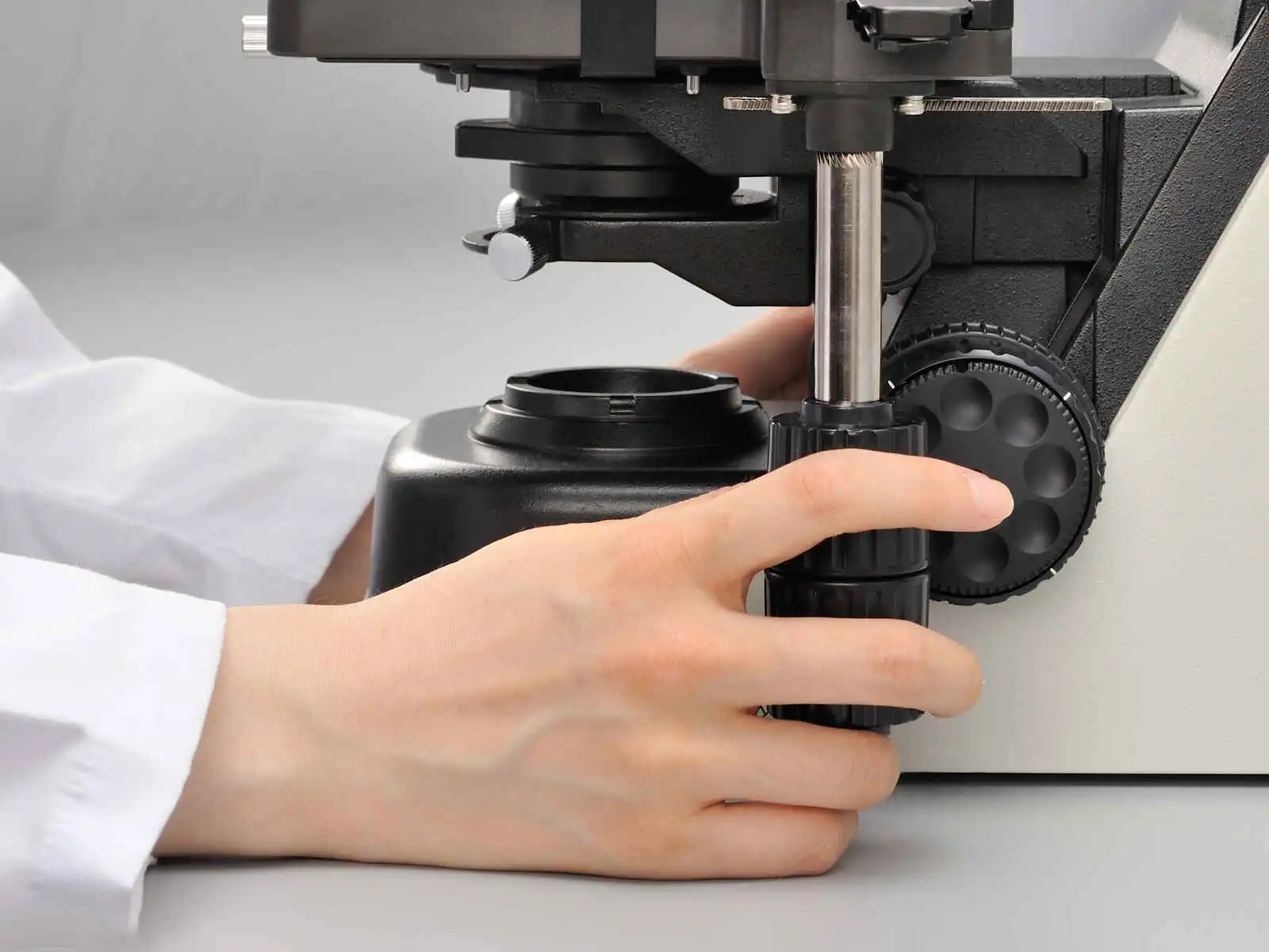
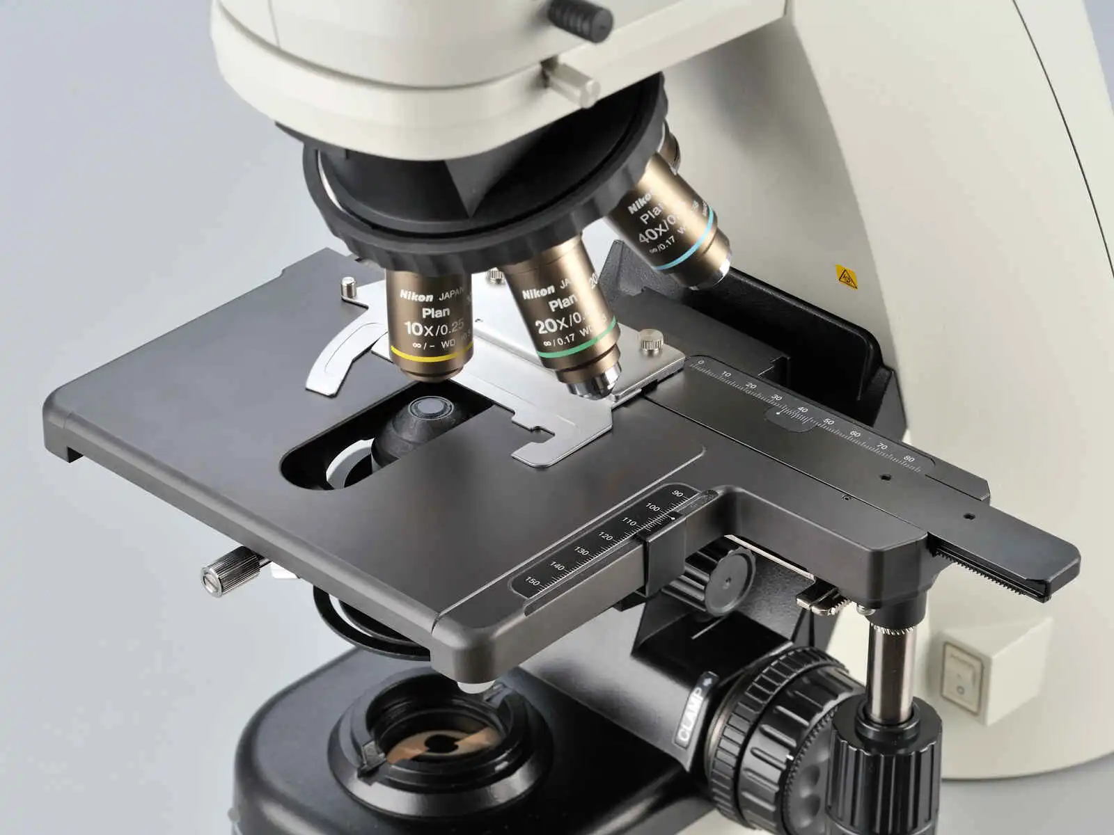
Without spacer (left), with spacer (right) Height adjustable stage handle Ceramic-coated stage
Effortless image capturing
One simple click of the image capture button on the microscope base during observation enables the Digital Sight camera to capture the specimen image. Camera control software for tablet PC NIS-Elements L is equipped with a scene mode that can automatically set the optimum shooting conditions for each observation method. Since it has a network function, you can share images with a remote PC.
* NIS-Elements L is not for clinical diagnostic use.

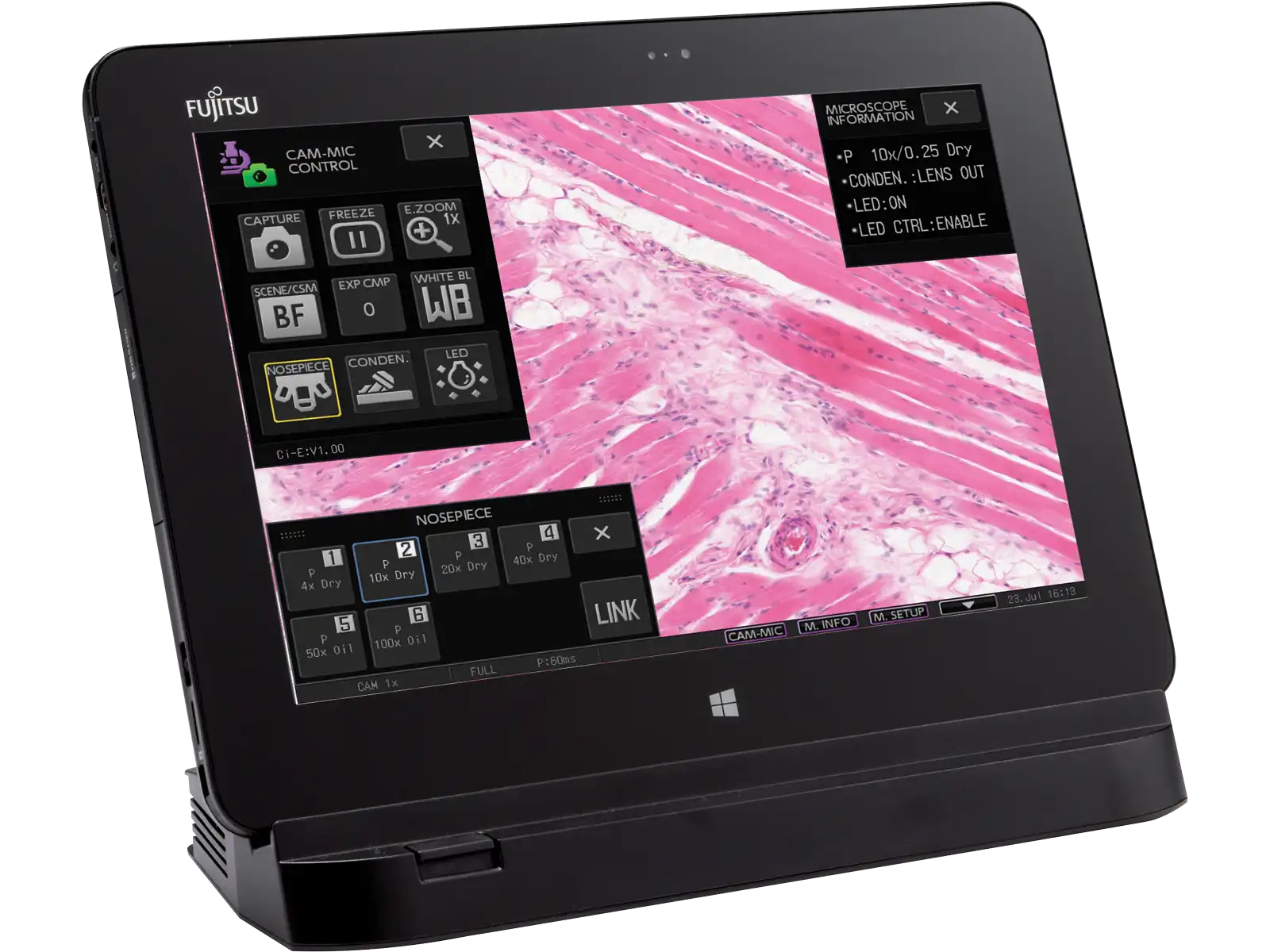
Image capture button NIS-Elements L camera control software for tablet PC
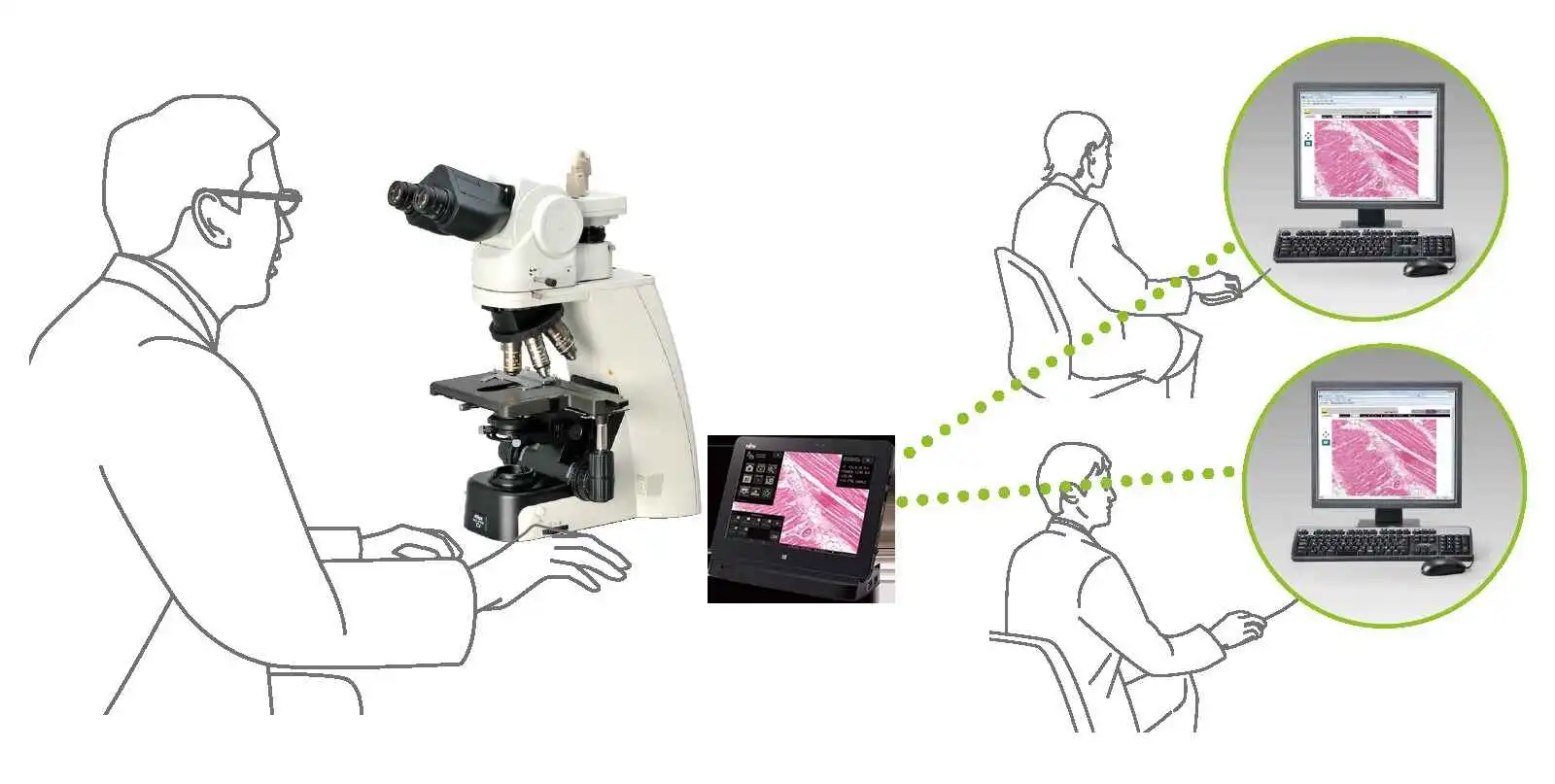
Digital pathology via network
Versatile observation techniquesPhase contrast
High-contrast images with neutral background coloration regardless of the magnification range can be captured. This observation technique is suitable for observation of unstained structures.

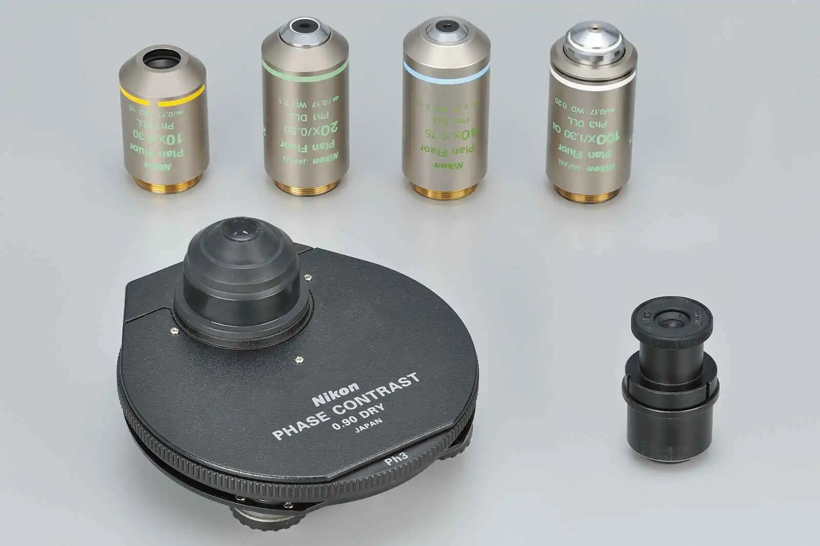
Phase contrast accessories and objective lenses
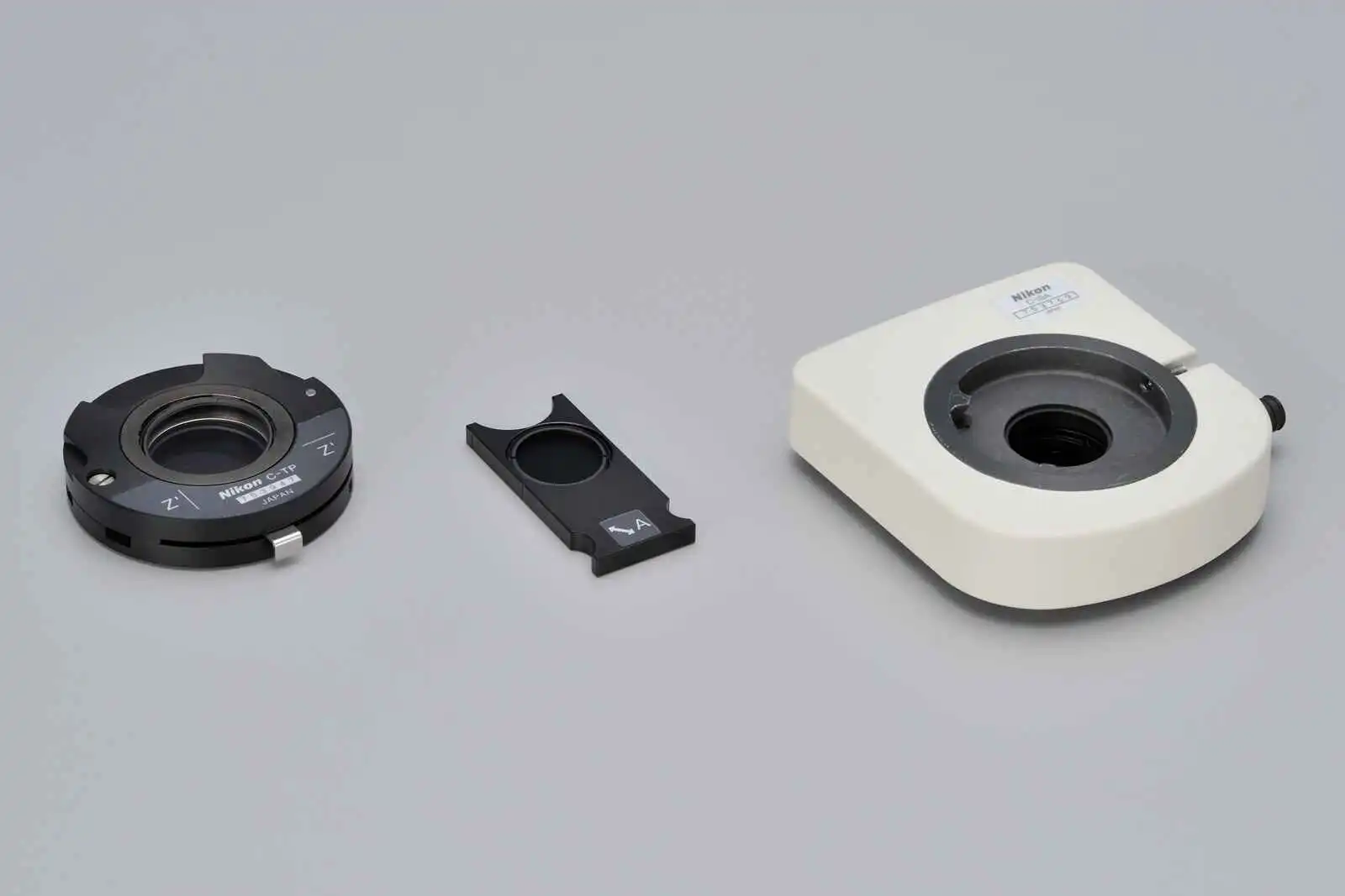
Simple polarizing
Ideal for observing bi-refringent samples such as collagen, amyloids and crystals.
* Two types of analyzer are available: intermediate tube type and nosepiece slider type.
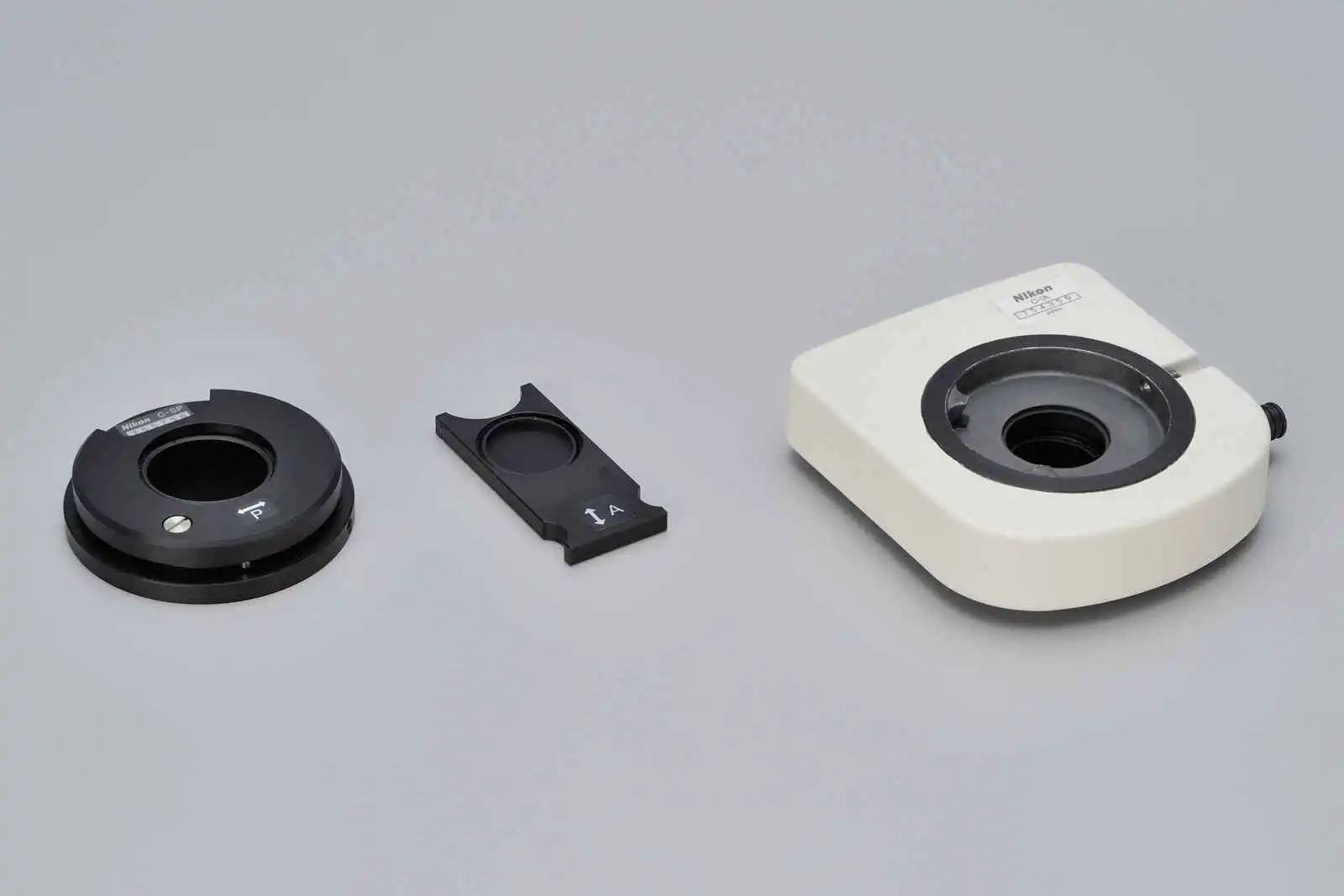
2.8-Dihydroxyadenine crystals, Simple polarizing accessories
Simple polarizing, CFI Plan Fluor 40X
Department of Clinical laboratory,
Nihon University Itabashi Hospital
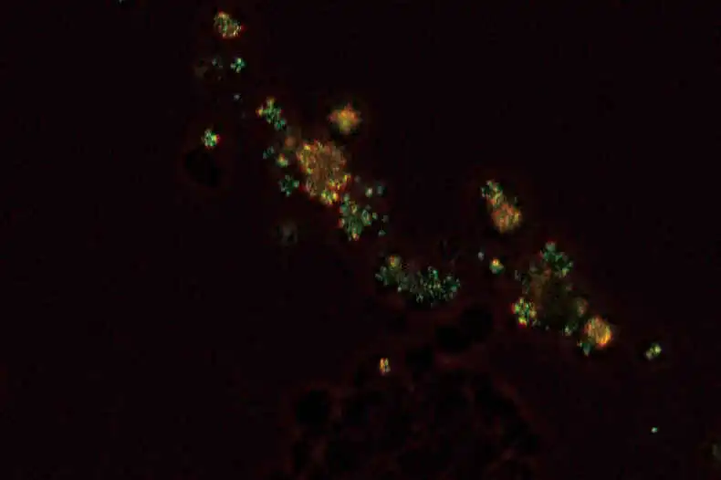
Sensitive color polarizing
Enables Identification of uric acid crystals by changes in the interference color. This technique is ideal for gout and pseudo-gout tests.
* Two types of analyzer are available: intermediate tube type and nosepiece slider type.
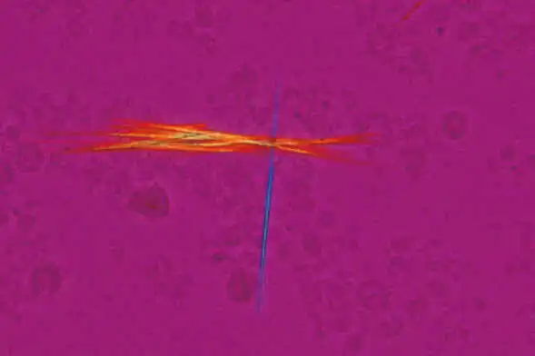
Sodium urate crystals, Sensitive color polarizing accessories
Sensitive color polarizing,CFI Plan Fluor 40X
Department of Clinical laboratory,
Nihon University Itabashi Hospital
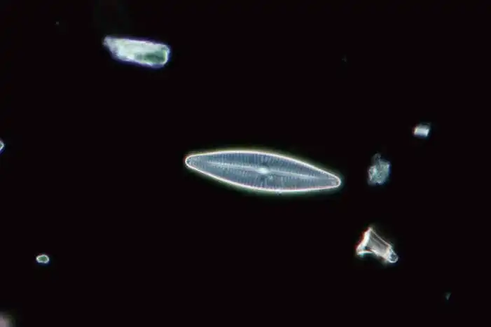
Darkfield
Enables clear observation of blood or minute structures such as flagella. Dry- and oil-type condensers are available. An expander lens is utilized for brighter imaging.
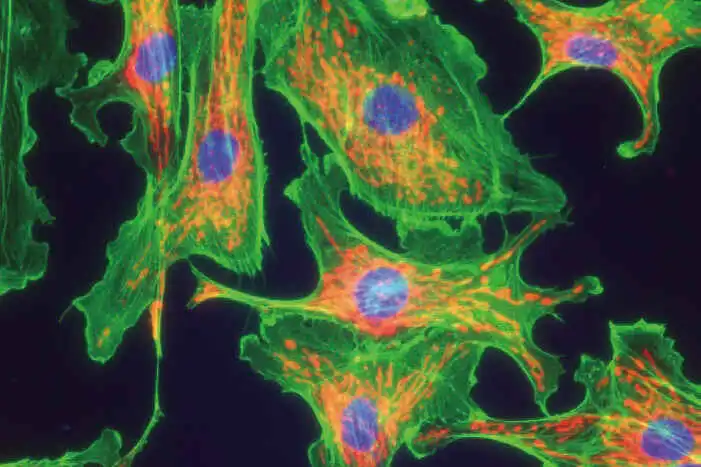
Epi-fluorescence
Compact epi-fluorescence attachments that utilize noise terminating mechanisms allow weakly fluorescing specimens to be captured with great clarity and brightness. Both CI-FL epi-fluorescence attachment (incorporates up to 4 filter cubes) and D-FL epi-fluorescence attachment (incorporates up to 6 filter cubes) allow easy switching of filter cubes. High-optical-performance objective lenses for epi-fluorescence imaging, including the CFI Plan Apochromat Lambda series and the CFI Plan Fluor series, are available.
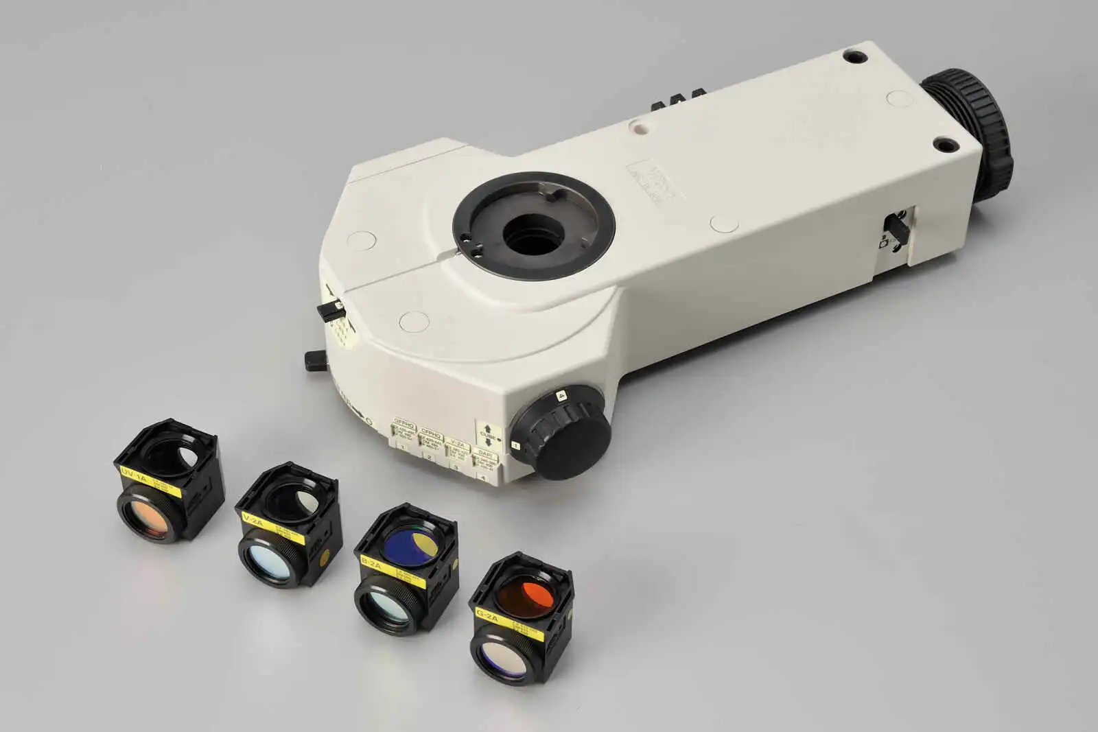
CI-FL epi-fluorescence attachment and filter cubes
| |
Ci-E |
| Optical system |
CFI60 Infinity Optical System |
| Illumination |
High luminescent White LED Illuminator (Eco-illumination) |
| Automatic intensity reproduction function |
| Controls |
Image capture button |
Nosepiece rotating buttons
Remote control pad |
Eyepieces
(F.O.V. mm) |
- CFI 10× (22)
- CFI 12.5× (16)
- CFI 15× (14.5)
- CFI UW 10× (25)
|
| Focusing |
Coaxial Coarse/Fine focusing, Focusing stroke: 30 mm, Coarse: 9.33 mm/rotation, Fine: 0.1 mm/rotation
Coarse motion torque adjustable, Refocusing function |
Tubes
F.O.V. 22 mm
(Eyepiece/Port) |
- C-TB Binocular Tube
- C-TE2 Ergonomic Binocular Tube (100/0, 50/50 via optional C-TEP2 DSC Port or C-TEPF2.5 DSC Port F2.5x)
Inclination angle: 10-30 degree, Extension: up to 40 mm |
Tubes
F.O.V. 25 mm
(Eyepiece/Port) |
- C-TF Trinocular Tube F (100/0, 0/100)
- C-TT Trinocular Tube T (100/0, 20/80, 0/100)
|
| Nosepieces |
- Motorized Sextuple Nosepiece with Analyzer Slot (Within main body)
Switching between two objectives function |
| Stages |
Cross travel 78 (X) × 54 (Y) mm, with vernier calibrations, stage handle height and torque adjustable for all stages
C-HIC Double Arm Specimen Holder is available as an option for the below three stages.
- C-SR2S Right Handle Stage with 2S Holder
- C-CSR1S Right Handle Ceramic-coated Stage with 1S Holder
- C-CSR Right Handle Ceramic-coated Stage (C-H2L Specimen Holder 2L and C-H1L Specimen Holder 1L can be attached)
|
Condensers (NA)
Motorized |
- CI-C-E Motorized Swing-out Condenser (0.9/0.22)
Focusing stroke: 27 mm |
Condensers
(NA)
Manual |
Focusing stroke: 27 mm
- C-AB Abbe Condenser (0.9)
- C-AR Achromat Condenser (0.8)
- C-DO Darkfield Condenser Oil (1.2-1.43)
- C-DD Darkfield Condenser Dry (0.8-0.95)
- C-PH Phase Contrast Turret Condenser (0.9)
- C-AA Achromat Aplanatic Condenser (1.4)
- C-SA Slide Achromat Condenser 2-100× (0.9)
- C-SW Swing-out Achromat Condenser 1-100× (0.9/0.11)
- C-SWA Swing-out Achromat Condenser 2-100× (0.9/0.22)
- C-LAR LWD Achromat Condenser (0.65)
|
| Observation methods* |
Brightfield, Epi-fluorescence, Darkfield, Phase contrast, Simple polarizing, Sensitive color polarizing |
| Epi-fluorescence attachment |
- CI-FL Epi-fluorescence Attachment (4 filter cubes mountable)
- D-FL Epi-fluorescence Attachmennt (6 filter cubes mountable)
ND4/ND8/ND16 filters, Noise Terminator mechanism |
| Epi-fluorescence light source |
- C-LEDFI Epi-Fl LED Illuminator
- C-HGFI/HGFIE HG Precentered Fiber Illuminator Intensilight (130W)
- Hg Lamphouse and Power Supply (100W)
|
| Power consumption |
13W (Brightfield configuration) |
| Weight (approx.) |
15.4 kg (Binocular standard set) |
* Observations except Brightfield require optional accessories.


 Microsystem
 Endoscopysystem
 Energysystem
 EndoscopyConsumables
 +86-21-54286005
 Room 602, Building 1, No. 111 Luxiang Road (Greenland Park Plaza), Baoshan District, Shanghai, China
  English
 中文
中文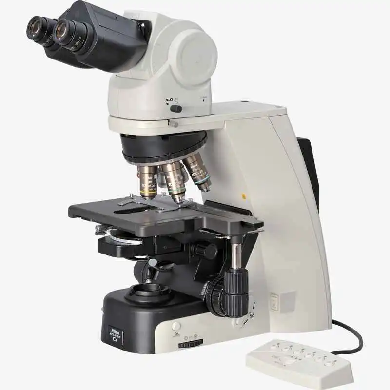
 ECLIPSE Ci-E_Brochure_EN


 中文
中文 English