 EndoscopyConsumables
 English
 中文
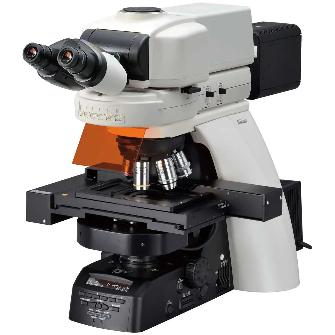
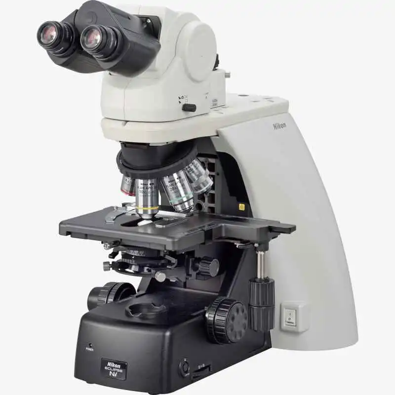
中文NIKON ECLIPSE Ci-S Upright Biological Microscope
Using the ergonomic binocular tube, which features an eyepiece that can be inclined from 10° to 30°and extended up to 40 mm, the microscope can be adjusted for natural posture.
Phone:+86-21-54286005


 Microsystem
 Endoscopysystem
 Energysystem
 +86-21-54286005
 info@tenmed.net
 Room 602, Building 1, No. 111 Luxiang Road (Greenland Park Plaza), Baoshan District, Shanghai, China

Using the ergonomic binocular tube, which features an eyepiece that can be inclined from 10° to 30°and extended up to 40 mm, the microscope can be adjusted for natural posture.
Phone:+86-21-54286005
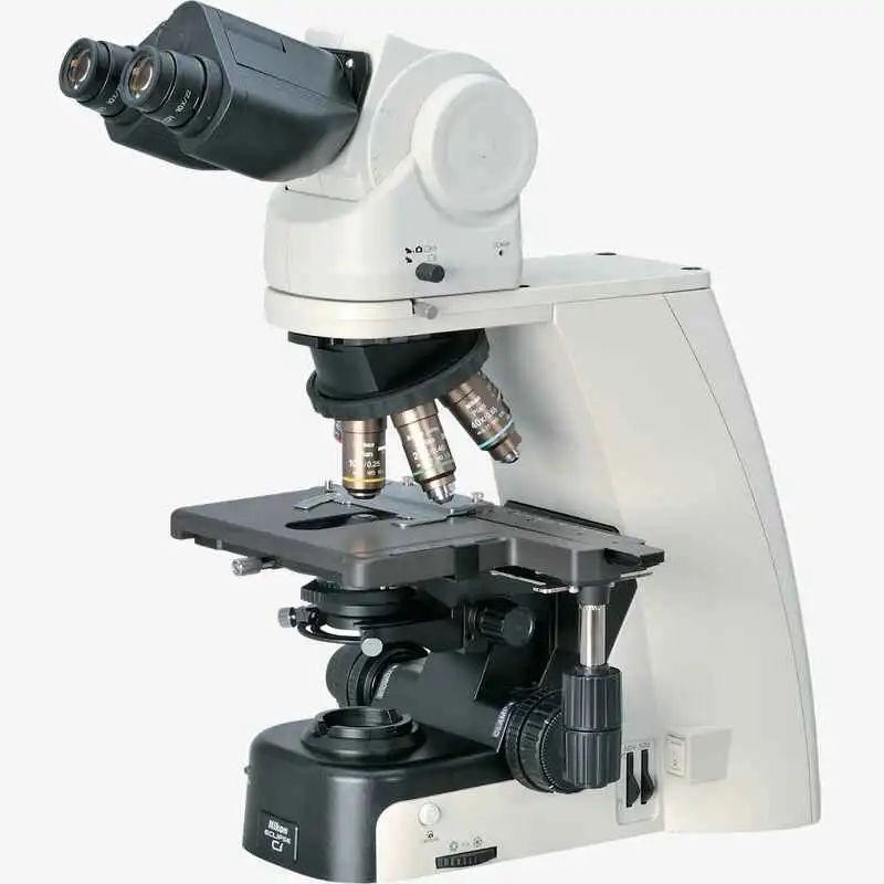
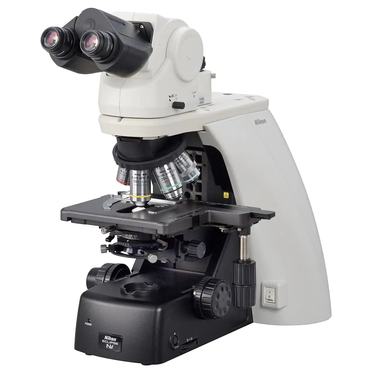
Using the ergonomic binocular tube, which features an eyepiece that can be inclined from 10° to 30°and extended up to 40 mm, the microscope can be adjusted for natural posture. The eyelevel riser lifts the eyepiece tube in 25 mm increments (up to 100 mm*) and can easily be adapted to suit users with different eye-point heights.
* Up to 50 mm with ergonomic binocular tube.

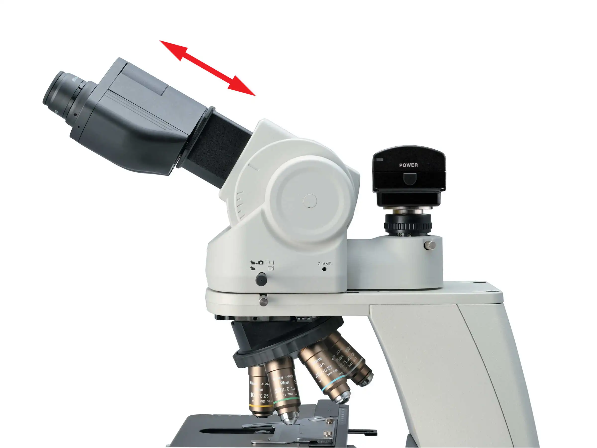
Ergonomic binocular tube Eyelevel riser
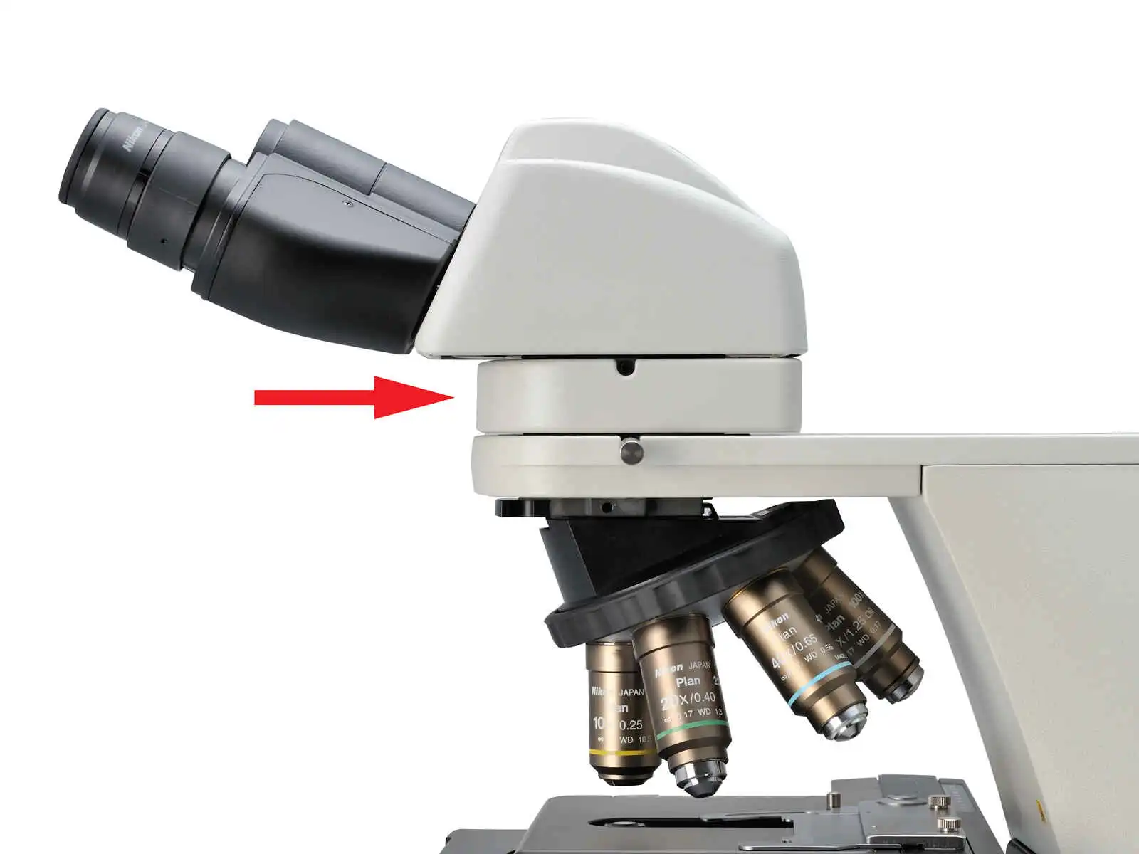
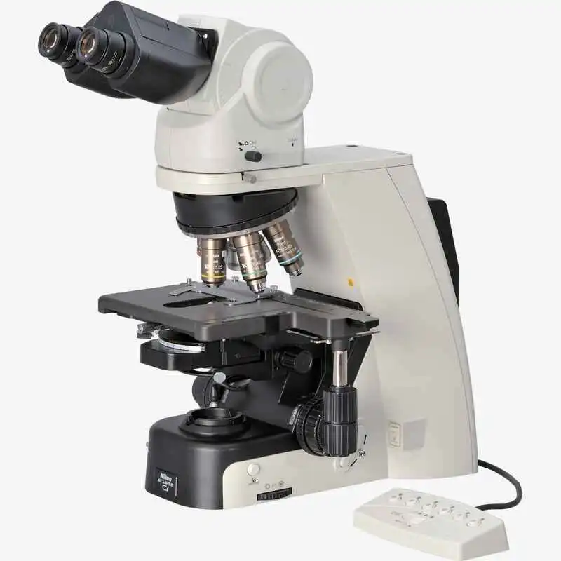
With the addition of a nosepiece spacer, the stage height can be lowered 20 mm from the standard position, reducing strain during frequent specimen change. The stage handle height can also be changed to ensure a comfortable hand position. The stage height can be locked using the refocusing knob, allowing quick refocusing after specimen changes. The stage is coated with a high-durability, scratch-resistant ceramic coating.
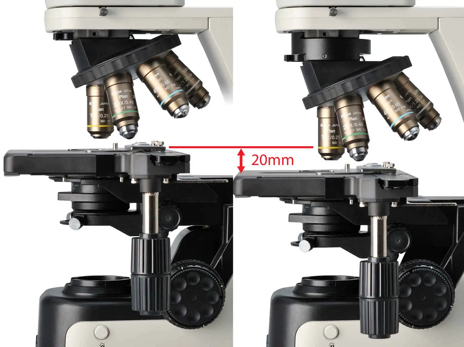
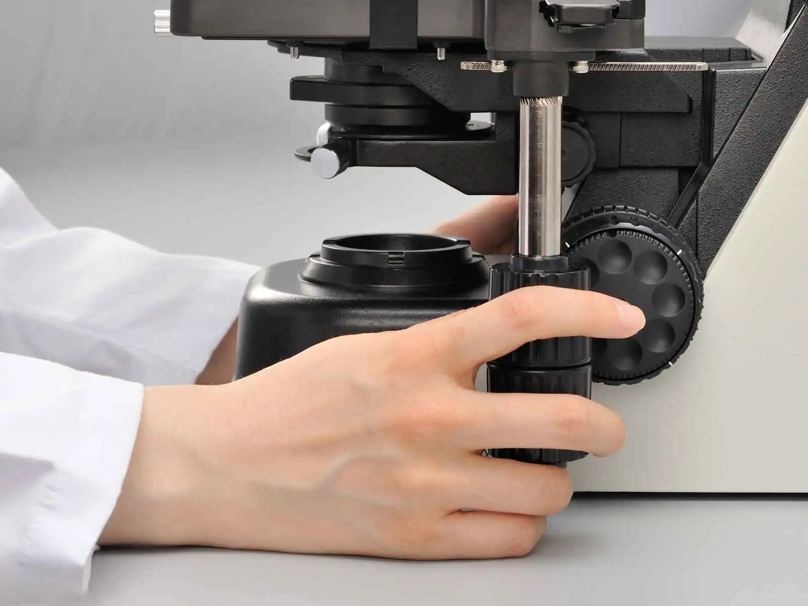
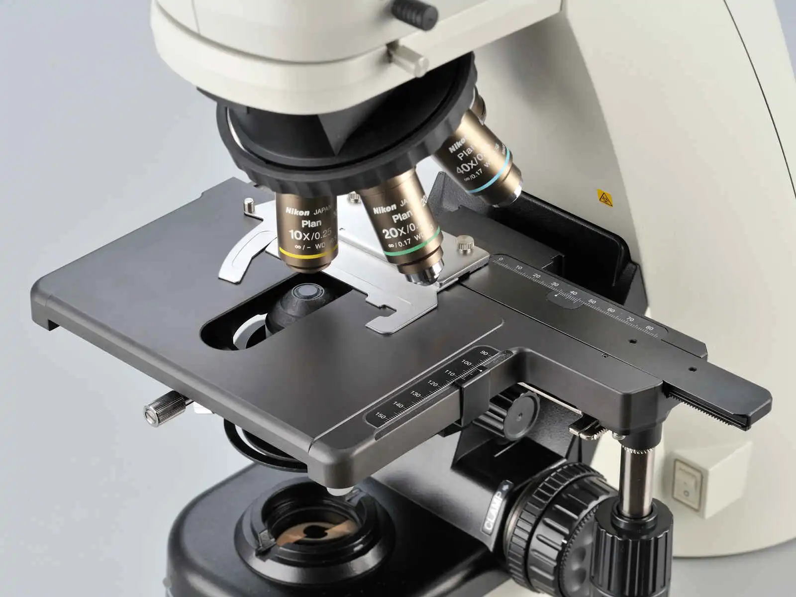
Without spacer (left), with spacer (right) Height adjustable stage handle Ceramic-coated stage
* NIS-Elements L is not for clinical diagnostic use.

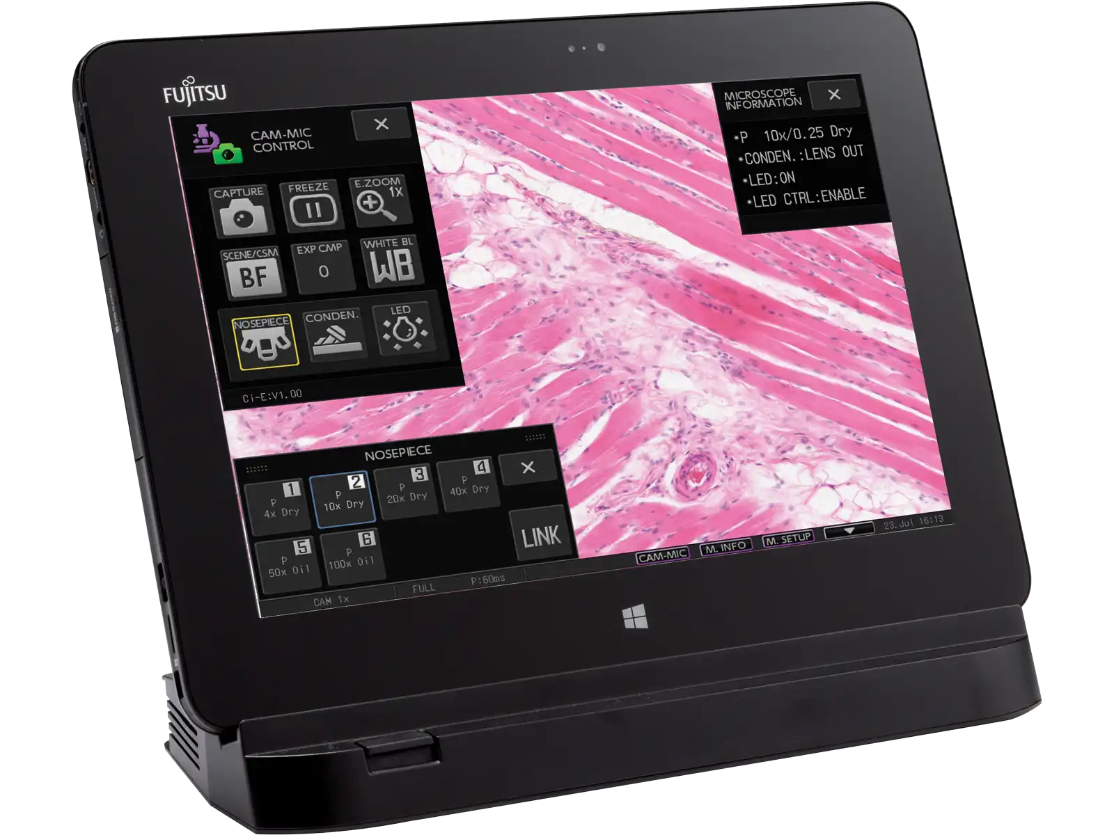
Image capture button NIS-Elements L camera control software for tablet PC
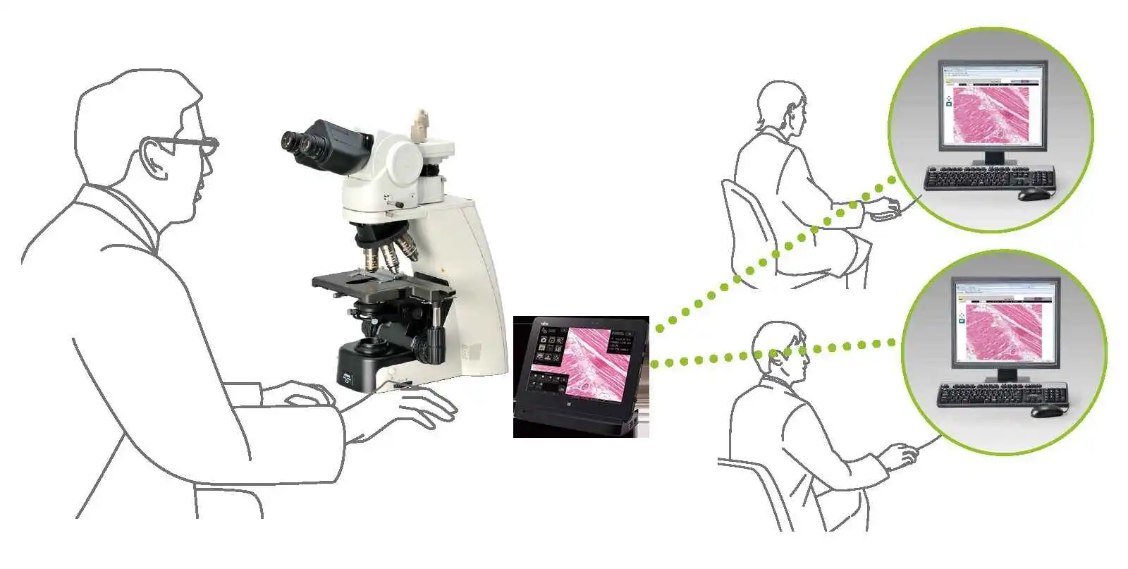
Digital pathology via network
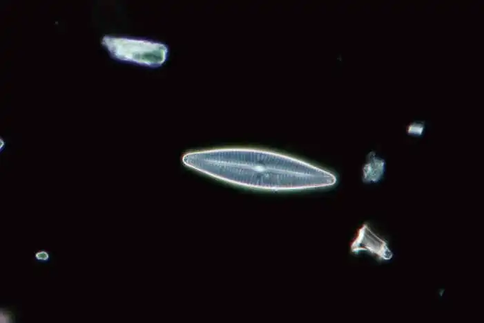
High-contrast images with neutral background coloration regardless of the magnification range can be captured. This observation technique is suitable for observation of unstained structures.

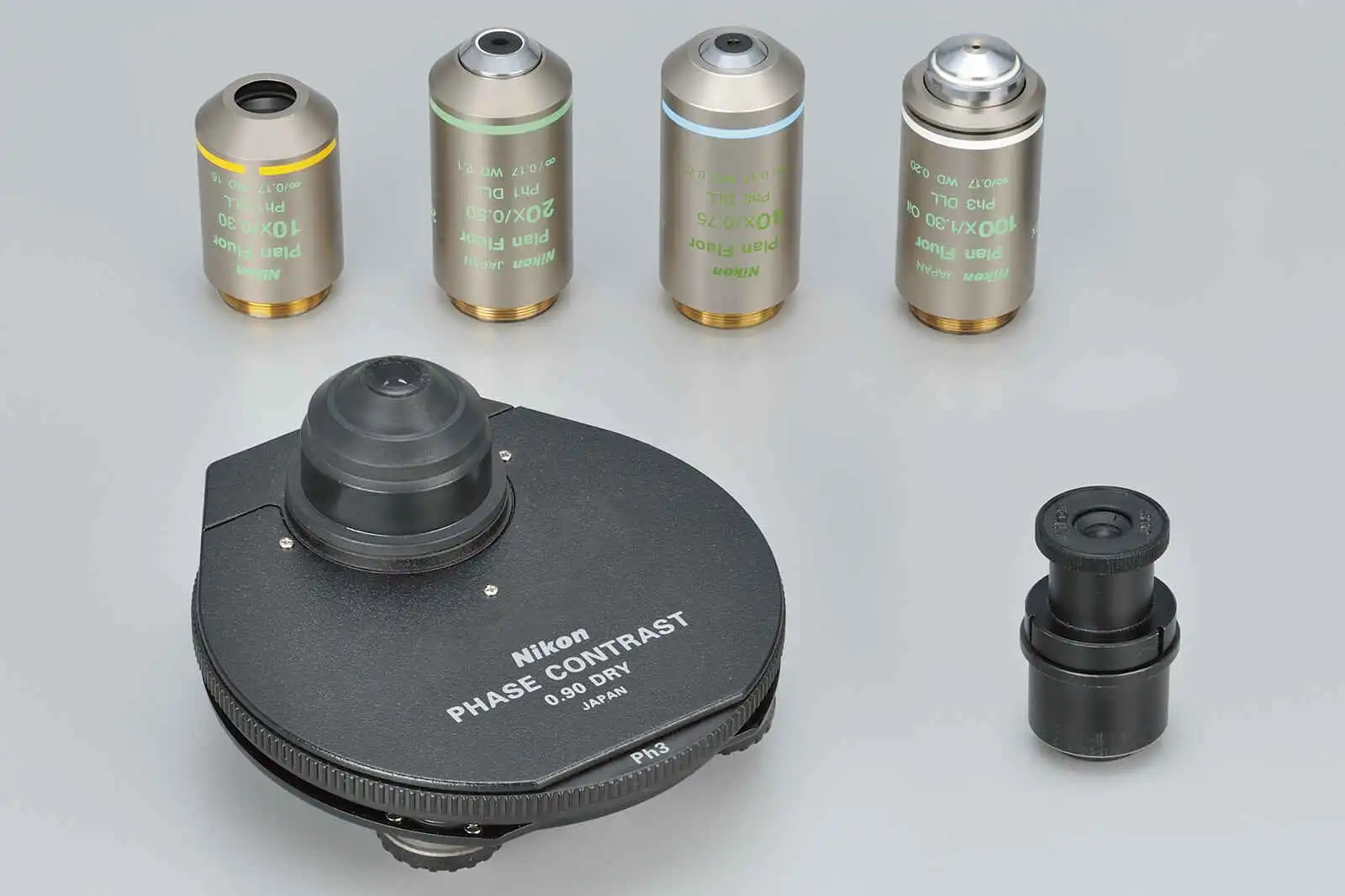
Phase contrast accessories and objective lenses
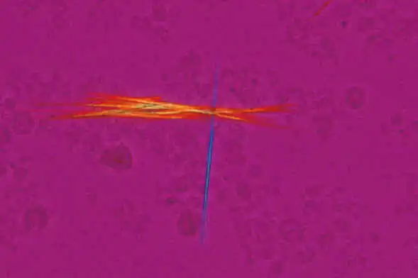
Ideal for observing bi-refringent samples such as collagen, amyloids and crystals.
* Two types of analyzer are available: intermediate tube type and nosepiece slider type.
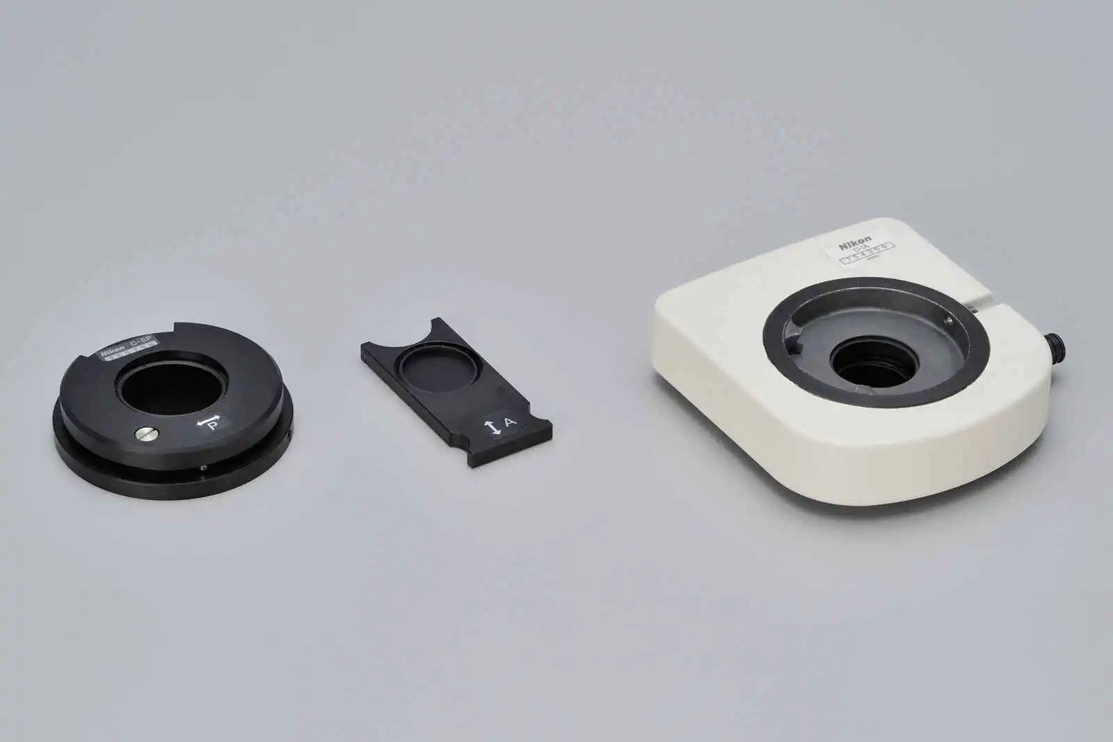
2.8-Dihydroxyadenine crystals, Simple polarizing accessories
Simple polarizing, CFI Plan Fluor 40X
Department of Clinical laboratory,
Nihon University Itabashi Hospital
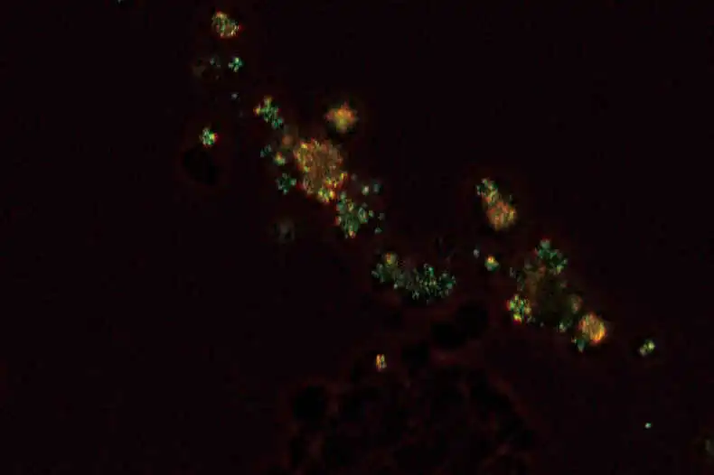
Enables Identification of uric acid crystals by changes in the interference color. This technique is ideal for gout and pseudo-gout tests.
* Two types of analyzer are available: intermediate tube type and nosepiece slider type.
Sodium urate crystals, Sensitive color polarizing accessories
Sensitive color polarizing,CFI Plan Fluor 40X
Department of Clinical laboratory,
Nihon University Itabashi Hospital
Enables clear observation of blood or minute structures such as flagella. Dry- and oil-type condensers are available. An expander lens is utilized for brighter imaging.
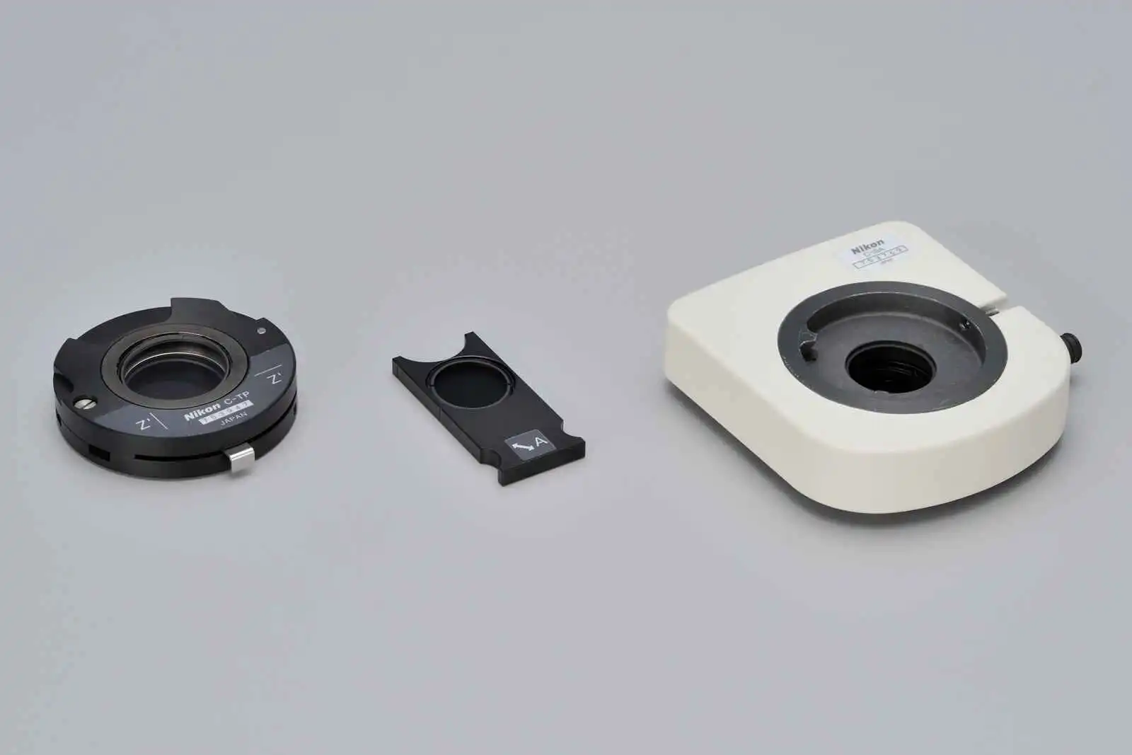
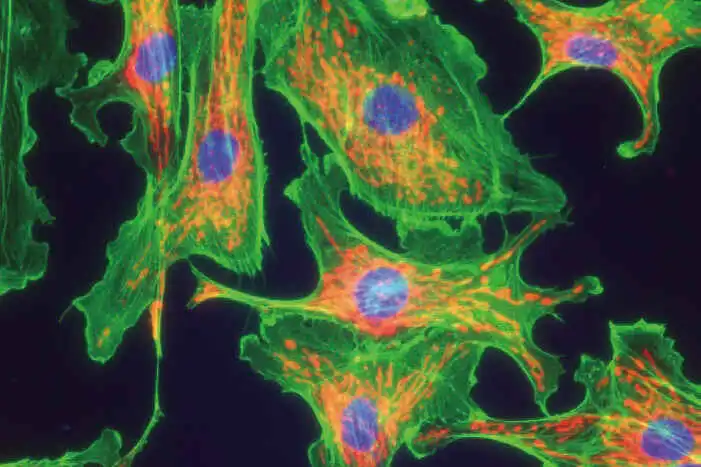
Compact epi-fluorescence attachments that utilize noise terminating mechanisms allow weakly fluorescing specimens to be captured with great clarity and brightness. Both CI-FL epi-fluorescence attachment (incorporates up to 4 filter cubes) and D-FL epi-fluorescence attachment (incorporates up to 6 filter cubes) allow easy switching of filter cubes. High-optical-performance objective lenses for epi-fluorescence imaging, including the CFI Plan Apochromat Lambda series and the CFI Plan Fluor series, are available.

CI-FL epi-fluorescence attachment and filter cubes


 中文
中文 English