 EndoscopyConsumables
 English
 中文
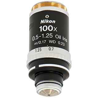
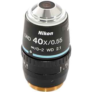
中文NIKON CFI Plan Fluor R-DS 40X Objectives NA 0.75 WD 0.66mm
NIKON CFI Plan Fluor R-DS 40X Objectives NA 0.75 WD 0.66mm
Phone:+86-21-54286005


 Microsystem
 Endoscopysystem
 Energysystem
 +86-21-54286005
 info@tenmed.net
 Room 602, Building 1, No. 111 Luxiang Road (Greenland Park Plaza), Baoshan District, Shanghai, China
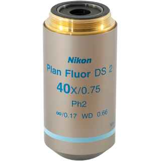
NIKON CFI Plan Fluor R-DS 40X Objectives NA 0.75 WD 0.66mm
Phone:+86-21-54286005
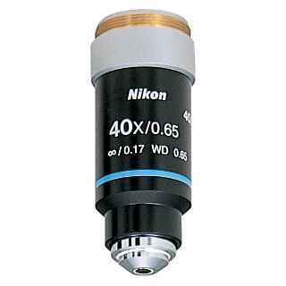
Dispersion staining (DS) microscopy refers to a family of analytical optical staining methods used to help determine the identity of unknown microscopic materials. The dispersive properties of the unknown material can be probed when immersed in a material with known dispersion curve. Color (wavelength) is then used as readout of differences in dispersive properties. DS microscopy is commonly applied in testing for the presence of asbestos in construction materials.
Nikon offers phase-type DS (R-DS) objectives to meet customer needs. R-DS phase contrast objectives for dispersion staining use a phase plate to block the waveband where the refractive indices of two materials are similar.


 中文
中文 English